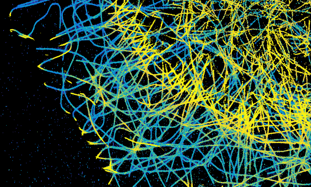
G-protein-coupled receptors mediate the biological effects of many hormones and neurotransmitters and are important pharmacological targets1. They transmit their signals to the cell interior by interacting with G proteins. However, it is unclear how receptors and G proteins meet, interact and couple. Here we analyse the concerted motion of G-protein-coupled receptors and G proteins on the plasma membrane and provide a quantitative model that reveals the key factors that underlie the high spatiotemporal complexity of their interactions. Using two-colour, single-molecule imaging we visualize interactions between individual receptors and G proteins at the surface of living cells. Under basal conditions, receptors and G proteins form activity-dependent complexes that last for around one second. Agonists specifically regulate the kinetics of receptor–G protein interactions, mainly by increasing their association rate. We find hot spots on the plasma membrane, at least partially defined by the cytoskeleton and clathrin-coated pits, in which receptors and G proteins are confined and preferentially couple. Imaging with the nanobody Nb37 suggests that signalling by G-protein-coupled receptors occurs preferentially at these hot spots. These findings shed new light on the dynamic interactions that control G-protein-coupled receptor signalling.


