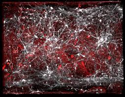
A snowflake begins life as a tiny crystal that acts as a seed on which water molecules aggregate, increasing the size of the snowflake as it descends to earth. Proteins can also act as seeds — for instance, in a class of age-related disorders called amyloid diseases, in which thousands of copies of a type of protein known as an amyloid adopt an abnormal structure and aggregate in harmful clumps. In Parkinson’s disease, aggregates of the amyloid protein α-synuclein accumulate in neurons. A rarer neurodegenerative disease, multiple system atrophy (MSA), involves α-synuclein aggregates in neuron-supporting cells called glia. It can be difficult to distinguish between the two disorders, given their overlapping symptoms, but they require different treatments. Writing in Nature, Shahnawaz et al.1 provide an explanation for this difference: like two dissimilar snowflakes composed of identical water molecules, α-synuclein aggregates form distinct 3D architectures in each disease.
In vitro and animal experiments have previously indicated that different aggregate structures of α-synuclein, called strains, yield different effects2. The various α-synuclein strains not only can have distinct cell-killing abilities and different seeding and propagation properties, but also can target different cell types and areas of the mammalian brain3,4.
Shahnawaz et al. built on these previous findings using a technique called protein misfolding cyclic amplification (PMCA), which amplifies small amounts of α-synuclein aggregate, allowing thorough examination of minuscule samples. An amyloid-specific fluorescent dye is incorporated into the newly formed aggregates, enabling their analysis.
Impressively, the authors amplified and analysed samples from the cerebrospinal fluid of more than 200 people who had either Parkinson’s disease or MSA, or who were healthy (Fig. 1). They found that samples taken from people with Parkinson’s disease displayed more fluorescence than those from people with MSA. Thus, PMCA could be used to discriminate between Parkinson’s disease and MSA.
The different levels of fluorescence suggested that the amyloid dye interacted with each α-synuclein aggregate differently, and that distinct α-synuclein strains are involved in the two diseases. The authors confirmed this result by showing that the two strains could also be distinguished by using proteinase K digestion (an enzymatic treatment that breaks down strains that have different structures in different ways), and through other biophysical characterizations, including a microscopy approach called cryo-electron tomography.
Shahnawaz and colleagues’ work has two major implications. First, it demonstrates that PMCA can be used as a diagnostic tool to discriminate between diseases involving α-synuclein. However, it should be noted that the samples analysed in this study were obtained from people who had already been diagnosed, and it remains unclear whether the approach could be used as a predictive tool to detect disease at earlier stages. Moreover, it is possible that PMCA is affected by the medication given to the participants who had Parkinson’s disease. These people typically receive the hormone dopamine (L-dopa), which has been shown to affect α-synuclein aggregation in vitro5.
Second, the study adds to a growing body of evidence supporting the ‘one polymorph, one disease’ hypothesis6–8, which states that different structural forms (polymorphs) of the same aggregated protein can cause distinct pathologies and symptoms. What might lead a protein to adopt different structures? In vitro, distinct fold structures can result from distinct environmental conditions. For example, different α-synuclein polymorphs arise depending on whether the protein is kept in a phosphate-containing or phosphate-free buffer9. In vivo, α-synuclein is exposed to several environments. Indeed, the neurons that degenerate in Parkinson’s disease and the glia affected in MSA belong to different cell lineages, and have markedly different intracellular environments. In addition, α-synuclein can move between cells, exposing it to both intra- and extracellular environments2.
The idea of different polymorphs in disease dates back to studies of prion proteins6 in the 1990s. Much like amyloids, prions aggregate in harmful infectious clumps to cause neurodegenerative conditions such as Creutzfeldt–Jakob disease in humans and scrapie in sheep. Several strains of prion, each adopting a different polymorph, typically coexist in a given sample or organism7. The strains have different fitnesses in different environments, which governs their ability to replicate7 — a phenomenon known as the prion cloud10.
A corollary of this idea is that if environmental conditions change, the relative abundance of each polymorph might change. This principle also governs the PMCA assay. Under given conditions, the fittest polymorphs should be amplified from a possible mix of pre-existing strains. Indeed, in Shahnawaz and colleagues’ experiments, a single distinct polymorph was amplified from Parkinson’s disease samples and another from MSA samples.
By contrast, in another recent study that used PMCA, Strohäker and colleagues11 reported no significant differences between structures of α-synuclein derived from the brains of people who had Parkinson’s disease and those with from people with MSA. A possible explanation for this apparent discrepancy is that the two groups used different PMCA protocols. In addition, Strohäker et al. used a much smaller group of patients than did Shahnawaz and colleagues. In fact, analysis using nuclear magnetic resonance spectroscopy did indicate distinct structural features in a subset of Strohäker and colleagues’ samples.
High-resolution cryo-electron microscopy has been used to demonstrate the existence of distinct disease-specific polymorphs of another neurodegeneration-associated protein, tau, at atomic resolution8. A similar approach using samples extracted under mild conditions might give us a clearer picture of the reality for α-synuclein. Taken together with similar observations for Alzheimer’s disease12, our understanding of the structural landscape of amyloid diseases is broadening.


