Highlights
- Modern neuroscience approaches have expanded investigation into the functional role of the claustrum, one of the most highly connected regions of the brain.
- Emerging data across rodent studies demonstrate that the claustrum is required for optimal cognitive performance and synchronizes distant cortical areas.
- Human whole-brain functional imaging data demonstrate the claustrum activates during difficult versions of a cognitive task and with the emergence of task-positive cortical networks.
- We propose a functional role for the claustrum in cortical network instantiation underlying cognitive control.
Early hypotheses of claustrum function were fueled by neuroanatomical data and yielded suggestions that the claustrum is involved in processes ranging from salience detection to multisensory integration for perceptual binding. While these hypotheses spurred useful investigations, incompatibilities inherent in these views must be reconciled to further conceptualize claustrum function amid a wealth of new data. Here, we review the varied models of claustrum function and synthesize them with developments in the field to produce a novel functional model: network instantiation in cognitive control (NICC). This model proposes that frontal cortices direct the claustrum to flexibly instantiate cortical networks to subserve cognitive control. We present literature support for this model and provide testable predictions arising from this conceptual framework.
A primer on the claustrum
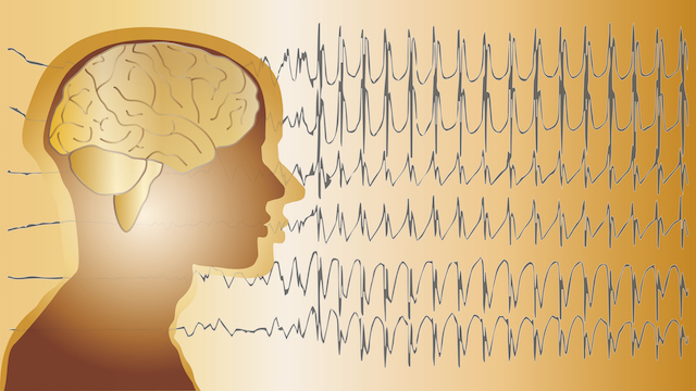
The claustrum is a thin, elongated subcortical telencephalic nucleus (Figure 1). It possesses bidirectional connections with many areas of the cerebral cortex and receives input from select subcortical regions [1., 2., 3., 4., 5., 6., 7., 8., 9., 10., 11., 12., 13.] (Figure 2 and Table S1 in the supplemental information online). The claustrum is positioned between the insula and putamen and, in primates, is bounded by the external and extreme capsules (Figure 1). The claustrum appears in all therian mammals, at least some monotremes [14., 15., 16.], and a potential homolog was recently discovered in reptiles [17.,18.] and possibly birds [19.]. The claustrum is composed of spiny glutamatergic projection neurons that exit the claustrum to predominantly target the cortex. In addition, multiple subpopulations of GABAergic, aspiny interneurons reside in the claustrum. They are differentiated by their expression of calcium-binding proteins or neuropeptides, including parvalbumin [20., 21., 22.], somatostatin, and vasoactive intestinal peptide [20.].
The elongated shape of the claustrum and its proximity to the dorsolateral striatum (putamen in primates), the insular cortex, and white matter structures have historically limited the ability to specifically manipulate the claustrum. As a result, most early hypotheses of claustrum function are disproportionately based on structural data obtained from neuronal tract-tracing. However, the advent of neural circuit-specific approaches and whole-brain functional imaging enables a more direct assessment of claustrum function through the recording of claustrum activity in the context of behavior and cognition. Considering the resulting expanded functional dataset, the diverse field of claustrum hypotheses may now be revisited and refined.
Here, we first describe previous hypotheses of claustrum function before advancing a new theory that unifies earlier concepts: specifically, that the claustrum receives frontal cortical signals for coordinating the engagement of downstream cortical areas to meet cognitive demands. We term this model NICC. We describe evidence for the NICC model, examine the model’s similarities and differences with other theories of claustrum function, and articulate predictions of the NICC model that can be tested in future research.
Existing functional hypotheses
Models of claustrum function fall into three ‘families’ of hypotheses that relate to: integration of cortical sensory information, salience and attentional processing, and cortical network dynamics. These hypotheses will be described in roughly the order they have been proposed.
Integration
Guided by an early claustrum lesion study in dogs in which auditory conditioning was impaired [23.], along with observations of non-somatotopic multimodal responses in the claustrum, Spector et al. [24.] hypothesized that the claustrum associates sensory stimuli across modalities. The involvement of the claustrum in sensory processing was supported in an earlier study in curarized cat in which claustrum neuronal firing was observed in response to somatic nerve stimulation [25.].
In an influential proposal, Ettlinger and Wilson [26.] postulated that all areas of the cortex have access to information from other (sensory) cortices through the claustrum, passing information through a series of cortico-claustro-cortical channels. In this way, the claustrum would function as a cross-modal transfer locus to support sensory integration. Crick and Koch [27.] expanded upon the Ettlinger and Wilson [26.] sensory integration model by proposing that the claustrum participates in perceptual binding by coordinating cortical processing. Perceptual binding refers to the process by which prior experience and ongoing sensory information gathered across multiple modalities are combined into a singular, cohesively perceived experience.
Following tract-tracing studies demonstrating widespread bidirectional claustrum connectivity with the cerebral cortex, Alloway et al. [28.] proposed a somewhat divergent function compared with Crick and Koch. They proposed a sensorimotor integration function that, at least in rodents, would serve to bilaterally coordinate whisker movement. This proposal was a landmark in proposing motor control as a possible outcome of claustrum-mediated sensory integration. Later, an investigation by Smith et al. [29.] established that the claustrum receives input from the whisker representation in motor cortex (but not somatosensory cortex). This led to an updated proposal that the claustrum cannot participate in cross-modal integration of whisker representations, though it may still participate in coordination of the two cortices. More recently, Chevee et al. [30.] detected few to no responses to sensory stimuli in mouse anterior claustrum, but instead observed abundant motor planning responses during a cross-modal sensory task.
In an elaboration upon the ‘binding hypothesis’ of Crick and Koch, Smythies et al. [31.] proposed that the claustrum synchronizes oscillations from different cortical regions to give rise to binding. This hypothesis was further refined by Smythies et al. [32.], who, similar to Crick and Koch [27.], speculated about the existence of intra-claustral interactions and the possibility of dynamic cortico-claustro-cortical circuits that would serve as a mechanism to regulate cortical synchrony for perceptual binding.
These iterations of the ‘binding hypothesis’ rely upon claustrum neurons being responsive to multimodal sensory inputs. While a function for the claustrum in multimodal sensory processing is supported by early work in curarized cat [24.,25.] and during multimodal tasks in human positron emission tomography [33.], it is challenged by later observations of unimodal responses to naturalistic stimuli in awake monkeys [34.] and an observed lack of sensory responses in anterior claustrum of mouse in response to visual or tactile stimuli [30.]. The ‘binding hypothesis’ also suggests that the claustrum is integral in producing states of consciousness. However, in a study of five epilepsy patients, bilateral electrical stimulation of the claustrum did not result in loss or alteration of subjective awareness [35.], and the claustrum remains relatively quiescent during a conscious resting state [24.,25.,34.,36.]. Despite a lack of strong evidence for the claustrum as a conductor of consciousness, these series of hypotheses generated core concepts that guided the emergence of a new generation of salience and attentional models of claustrum function.
Salience detection and/or attention
The salience detection/attention model of claustrum function was prompted by anatomical data showing that the claustrum is strongly connected with the anterior cingulate cortex (ACC), which is implicated in monitoring conflicts in decision-making processing [37.,38.] and is a component of the salience network (SN) [39.,40.]. In this model, as proposed by Remedios et al. [41.], the claustrum identifies changes in the sensory scene to alert other brain areas, a process that was proposed to contribute to sensory awareness. Observations that responses in primate claustrum are primarily unimodal [34.], similar regardless of the type of auditory stimuli (naturalistic or conspecific vocalization), only active at stimulus onset, and scaled with signal-to-noise ratio [41.] support this hypothesis.
In an updated view, Goll et al. [42.] proposed that the claustrum mediates the allocation of cortical processing for selective attention. In this model, claustro-cortical projections suppress cortical activity to reduce processing of unattended information. Thus, the claustrum is proposed as an ‘attentional searchlight’, a term Crick [43.] coined when discussing a similar function for the thalamic reticular nucleus. This hypothesis was recently tested in mouse, where it was found that claustrum neurons in rodent appear to primarily encode signals related to motor planning, rather than the attended sensory modality [30.].
Later, in response to fMRI observations of functional connectivity between the claustrum, insula, medial prefrontal cortex, and cingulate cortex in rodent, Smith et al. [44.] proposed that the claustrum integrates sensory and limbic information to facilitate both bottom-up and top-down attentional processes. Specifically, the claustrum was proposed to integrate input from the basolateral amygdala, mediodorsal thalamus, and the medial prefrontal cortex to then distribute that information to cortical areas of the relevant modality, such as the frontal eye field, ultimately resulting in attentional and behavioral orientation to a stimulus; a function not unlike that proposed for the superior colliculus [45.]. Smith et al. [44.] proposed this model as a circuit-based elaboration of a previous hypothesis, forwarded by Reser et al. [6.], who suggested that due to the large voxel size in fMRI, some activity historically attributed to the anterior insula may be a product of claustrum activity. Thus, it would follow that some roles attributed to anterior insula (e.g., participation in the SN and salience processing) may in fact be claustrum functions. Jackson et al. [46.], in a recent review of claustrum literature, reinforced a focus on claustrum as a contributor to attentional processes by distributing limbic information to cortex to inform actions and perception via inhibitory control of cortex.
Cortical networks and cognitive control
The claustrum was recently proposed to play a role in cortical network switching [6.,7.]. In this section we will first give a brief overview of cortical networks and cognitive control, then more specifically describe how the claustrum fulfills a key mechanism of network interaction facilitating cognitive control. This lays the groundwork for the presentation of our new model of claustrum as a key hub modulating networks of cognitive control, presented in the next section.
Cognitive control is the ability to initiate, maintain, and monitor relevant information during goal-directed action, as opposed to habitual stimulus-response behavior [47.,48.]. Cognitive control, therefore, enables adaptive behavior in response to environmental challenges. This is distinct from other cognitive subprocesses such as selective attention, which is the ability to increase signal-to-noise detection of particular sensory modalities over others or to increase signal-to-noise detection of features within a sensory modality [49.]. As an umbrella cognitive faculty, cognitive control is the ability to muster, for instance, working memory and selective attention processes for task performance.
The degree to which cognitive control is implemented, known as cognitive demand, is a function of task difficulty [50.]. Task difficulty increases with task variable and rule number, as well as rule complexity. In an animal laboratory setting, an example of a ‘cognitively nondemanding’ task would be a requirement to nose-poke a lone port each time it illuminates for a sugar reward (e.g., the one-choice serial response time task). This contrasts with a ‘high cognitive demand’ rodent task wherein five ports may each, randomly, illuminate with each trial (e.g., the five-choice serial response time task), requiring the animal to monitor five different ports to successfully poke the one that illuminates. Despite the same physical requirement between the two tasks (correct nose-poke required for a reward), the five-choice task is more difficult to learn and, once learned, error rates are higher compared with the one-choice task version. Thus, the higher cognitive demand of the five-choice task requires greater mobilization of cognitive control to perform.
Cortical networks underlie cognitive control
Cognitive control demands flexible engagement of cognitive processes from moment to moment. Meeting these demands is the flexible engagement of networks of brain regions. Networks are defined as collections of functionally correlated regions (as assessed during whole-brain neuroimaging) identified during task-based or task-free (i.e., resting state) periods of measurement [51., 52., 53.] and largely include cortical regions [54., 55., 56.] across species [54.,57., 58., 59., 60., 61., 62., 63., 64.] (Box 1). Several groups attempted to map the human brain’s network organization [65., 66., 67., 68., 69.]. Such parcellations are not identical, but they consistently include local sensory and somatomotor networks, as well as reproducible, geographically distributed networks of association cortices. A widely cited seven-network parcellation [65.] of the human brain includes: visual and somatomotor networks; a default mode network (DMN) notable for increased metabolic activity at rest and decreased blood oxygen level-dependent signal during externally focused tasks [53.,70.]; a frontoparietal network (FPN) active during tasks; dorsal (DAN) and ventral (VAN) attention networks active during tasks; and a limbic network. More granular parcellations reveal additional networks, such as subcomponents of the DMN [54.], as well as the SN and a cingulo-opercular network (CON) overlapping with the VAN [52.]. More broadly, a commonly used gross taxonomy divides the brain into two anti-correlated networks: a task-positive network active during tasks and a task-negative network (the latter largely overlaps with the DMN) [71.].


