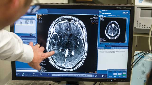
In cerebral amyloid angiopathy (CAA) and Alzheimer’s disease, insoluble aggregates of a peptide known as amyloid-β (Aβ) progressively build up in the spaces between cells, forming amyloid deposits. In Alzheimer’s disease, these aggregates are found between neurons, whereas in CAA, a related but not always coexisting condition, they are found in the walls of brain blood vessels. Aβ aggregates are thought to be early drivers of the pathological processes of CAA and Alzheimer’s disease that culminate in neurodegeneration. In 2015, researchers reported evidence of early Aβ pathology in the brains of some people with growth deficiency who had been treated with human growth hormone collected from pituitary glands at autopsy1. This finding raised the possibility that Aβ pathology might be transmissible between humans under certain conditions through contaminated brain-tissue derivatives. Writing in Nature, Purro et al.2 provide further support for this hypothesis.
From 1958 to 1985, approximately 30,000 children with growth deficiency were treated with cadaver-derived growth hormone (c-hGH) worldwide3. In 1985, three recipients were found to have developed Creutzfeldt–Jakob disease (CJD), which is fatal. CJD belongs to a group of diseases known as transmissible spongiform encephalopathies, which are characterized by progressive and irreversible brain damage resulting from the accumulation of a misfolded form of a brain protein called prion protein. These abnormal prion proteins can themselves cause normal prion proteins to misfold, and thus spread the disease. Given the evidence that contaminated c-hGH had caused CJD, this type of treatment was quickly stopped and synthetic recombinant human growth hormone (rhGH) became the standard of care.
Alzheimer’s disease is not a classic prion disease, but shares characteristics with this type of disorder. Misfolded, aggregated Aβ peptides and tau proteins, which are toxic to neurons, are present in the brain as key components of Alzheimer’s disease. Inoculation of minute amounts of misfolded Aβ (known as Aβ ‘seeds’) isolated from the brains of people with Alzheimer’s disease can induce build-up of Aβ deposits (called Aβ plaques) in non-human primates4, and brain extracts from people or mice that develop Aβ plaques can also cause accelerated plaque accumulation when given to genetically modified mice5.
The 2015 finding of Aβ plaques and CAA in the brains of seven of eight recipients of c-hGH therapy who had died of CJD further supported the idea that Aβ pathology can be transmitted through a prion-like mechanism1 (Fig. 1). Aβ pathology is rarely found in young adults without genetic risk factors for Alzheimer’s disease or CAA, so the findings suggested that the c-hGH used to treat the patients might have been contaminated with Aβ seeds in addition to misfolded prion proteins.
To provide more-direct evidence that the Aβ deposits found in these people resulted from Aβ-seed contamination, Purro and colleagues first tested whether Aβ was present in vials of c-hGH from batches that had been used to treat patients and that had been stored since the 1980s. Growth hormone is produced in the pituitary gland, a small structure found at the base of the brain. To obtain c-hGH, the pituitary glands from thousands of donors had been pooled and mixed, and the hormone had been chemically extracted using various preparation methods. Patients received c-hGH from multiple batches. However, all of those who were treated in the United Kingdom and developed CJD — 38 people by the year 2000 (ref. 6)6 — received injections from batches prepared using a method called the Hartree-modified Wilhelmi procedure (HWP).
Purro et al. detected Aβ in all c-hGH samples prepared using the HWP method, but not in those prepared using any of three other methods. Size-exclusion chromatography, a separation technique used in all non-HWP preparations, might have reduced contamination by Aβ peptides.
The authors went on to show that these HWP preparations of c-hGH possess Aβ-seeding ability by injecting them into mice genetically engineered to express human versions of Aβ (Fig. 1). Mice inoculated with HWP-prepared c-hGH developed markedly more Aβ plaques and CAA than did those inoculated with synthetic rhGH.
These results provide strong evidence that the Aβ pathology previously reported in people who died of CJD after receiving c-hGH1 was indeed caused by their treatment. The data also corroborate previous studies in genetically modified mice demonstrating that misfolded Aβ can behave in a prion-like fashion5. Future studies should investigate the amount of Aβ these patients received over their treatment course, to try to determine the threshold of misfolded Aβ concentration required to transmit Aβ-plaque formation or CAA.
The c-hGH preparations shown in this study to induce Aβ pathology in mouse brains were injected directly into the brain, whereas the affected humans had received injections through other routes (intravenously or intramuscularly). Future studies in animals should assess whether the route of administration influences the ability of material containing misfolded Aβ to cause brain Aβ pathology, and should investigate the minimum amount of material that has pathological effects.
The eight people with therapy-induced CJD who, with one exception, also had Aβ pathology had an incubation period from their last c-hGH treatment to CJD onset of 18.8–30.8 years1. Aβ accumulation in people who develop dementia due to Alzheimer’s disease is estimated to precede disease symptoms by 15–20 years7,8. The seven people who developed Aβ pathology did not meet the full pathological criteria of Alzheimer’s disease, and whether they would have developed the clinical manifestations of the disease had they not died of CJD is unclear.
Purro et al.2 report that the HWP c-hGH batches also contained misfolded tau proteins. This study did not show evidence of tau-pathology transmission, and the earlier study of people with therapy-induced CJD did not detect misfolded tau proteins in their brains1. Nevertheless, surveillance of surviving c-hGH recipients should continue to watch out for this possibility. Overall, treatment-related transmission of various brain pathologies cannot be ruled out.
Lastly, it is worth noting that the stored vials of c-hGH had been maintained at ambient temperature since the mid-1980s. Their ability to transmit Aβ pathology seen in this study corroborates the idea that Aβ seeds are remarkably stable9. This property of Aβ seeds emphasizes the importance of not using biological material prepared from the human central nervous system for injection or transplantation into patients during neurosurgical or medical procedures, unless these materials are adequately screened or there is no other option. Similarly, it is crucial that surgical instruments that come into contact with the human brain are appropriately treated to remove seeds of misfolded forms of peptides and proteins such as Aβ, tau or prion protein.


