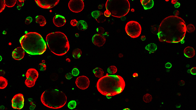
Highlights
- •Xenotransplanted brain organoids as in vivo platform for studying human microglia (hMGs)
- •hMGs gain human-specific transcriptomic signatures and assume in-vivo-like identities
- •hMGs engage in surveilling the human brain environment and react to perturbations
- •A patient-derived model reveals a brain-environment-induced immune response in autism
Summary
Microglia are specialized brain-resident macrophages that play crucial roles in brain development, homeostasis, and disease. However, until now, the ability to model interactions between the human brain environment and microglia has been severely limited. To overcome these limitations, we developed an in vivo xenotransplantation approach that allows us to study functionally mature human microglia (hMGs) that operate within a physiologically relevant, vascularized immunocompetent human brain organoid (iHBO) model. Our data show that organoid-resident hMGs gain human-specific transcriptomic signatures that closely resemble their in vivo counterparts. In vivo two-photon imaging reveals that hMGs actively engage in surveilling the human brain environment, react to local injuries, and respond to systemic inflammatory cues. Finally, we demonstrate that the transplanted iHBOs developed here offer the unprecedented opportunity to study functional human microglia phenotypes in health and disease and provide experimental evidence for a brain-environment-induced immune response in a patient-specific model of autism with macrocephaly.
Introduction
A specialized population of tissue-resident macrophages known as microglia play a central role in brain development, homeostasis, and tissue repair. Microglia develop from yolk-sac-derived erythromyeloid progenitors (EMPs) and are presumed to enter the brain between 4.5 and 5.5 gestational weeks in humans; they use tangential and radial migration routes through the ventricle or the mantle zone.
Mounting evidence from human and animal studies suggests that microglia may be implicated in various brain disorders, including neurodevelopmental conditions such as autism spectrum disorder (ASD).
However, until now, the ability to model interactions between human brain cells and microglia has been severely limited. To understand and study human microglial function in health and disease, novel platforms are needed that feature functionally mature cells operating within a physiologically relevant human brain environment, a critical step for modeling the cellular and environmental complexity that emerges from a cooperative interaction between the different cell types in the developing human brain.
The brain environment is instrumental in sustaining and orchestrating microglial identity. Two-dimensional (2D) cultured microglia lack a brain environment and assume a non-physiological, partially activated state, which is reflected in a dramatic shift in their gene expression profile and epigenetic landscape. Brain organoids, on the other hand, recapitulate some features of the brain’s three-dimensional (3D) structure, allow more organized tissue formation, contain multiple cell types, and are considered to be more mature than a 2D culture system. However, current models generated through guided approaches still lack cell types that are of non-ectodermal origin, such as microglia. Recent advances have allowed the generation of induced microglia-like cells from human pluripotent stem cells (hPSCs) in isolation, including human embryonic stem cells (hESCs) and induced pluripotent stem cells (iPSCs).
Short-term co-culture experiments have demonstrated that the incorporation of hPSC-derived microglia-like cells into organoids is feasible, but the extent to which these in vitro structures support maturation and long-term survival and whether the integrated cells ultimately acquire a state that fully resembles the identity of their in vivo counterparts remain unknown. Recent attempts to reinstate microglial identity were achieved through xenotransplantation of hPSC-derived microglia into transgenic immunocompromised mouse models. However, these approaches lack the ability to assess the interaction of human microglia (hMGs) with their human neuronal environment, which is crucial for studying the cooperative contribution of the two components in the context of human diseases.
To model microglia identity and to assess the interaction and response of hMGs to the human brain environment, we harnessed our recently developed xenotransplantation approach to develop a transplanted immunocompetent human brain organoid (iHBO) model that allows the investigation of hPSC-derived human microglia within vascularized human brain organoids under physiological conditions in vivo. The hMGs populate the human organoid graft, express microglia-specific markers, and show morphological features indicative of a resting and surveillance state. Using longitudinal transcriptomic profiling, we show that the hMGs in our system follow a stepwise developmental program toward acquisition of an immune-sensing microglial state, similar to what has been reported in studies on human brain tissue.
We demonstrate that the hMGs assume transcriptomic identities that closely resemble their counterparts in vivo. By contrasting transcriptomic profiles gained under human and mouse brain environmental conditions, our system also reveals that some of the previously reported hMG-specific patterns of gene expression are in part instructed by the human brain environment. Finally, our approach allows the functional assessment of hMG phenotypes within a human-brain-like environment under physiological and pathological conditions in vivo. Using two-photon microscopy within the vascularized human organoid graft, we demonstrate that hMGs actively engage in surveilling the human-brain-like environment and are capable of reacting to environment-induced injury. We demonstrate that our in vivo approach is suitable for dissecting hMG phenotypes during development and disease, and we provide evidence for a brain-environment-induced immune response in a patient-specific model of autism with macrocephaly. Thus, the system developed here provides an opportunity to study functional human-specific microglia phenotypes in health and disease.
Results
EMPs efficiently populate developing cortical organoids
We sought to establish a novel brain organoid model to recapitulate the colonization of the developing human brain through EMPs. In humans, yolk-sac-derived EMPs accumulate in the superficial marginal zone of the telencephalic wall and are presumed to enter the telencephalon from the pial surface between gestational weeks 4.5 and 5.5.
To model these early events of microglial entry into the human telencephalon, we used a recently developed protocol to obtain viable and pure populations of CD43+ EMPs. Generating hPSC lines constitutively expressing tdTomato (tdT) or GFP under the control of a chicken-beta-actin promoter (CAG:tdT, CAG::GFP) enabled us to assess the colonization of cortical organoids through EMPs (Figure S1, Figure S10A–S1F). Live imaging of tdT+ EMPs co-cultured with forebrain-specific cortical organoids revealed a highly efficient invasion of EMPs within 12 h (Video S1). Subsequent tissue clearing confirmed the presence of tdT+ EMPs that invaded the cortical organoid tissue (Video S2), in line with previous studies that showed the feasibility of incorporating induced microglia-like cells into organoids…


