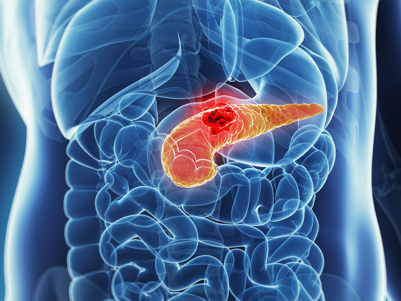
Significance
Carcinomas resist immunotherapy because T cells are absent from nests of cancer cells. The chemokine/chemokine receptor system, which regulates the migration of immune cells, is a candidate for this impaired intratumoral accumulation of T cells. Cancer cells in human pancreatic, colorectal, and breast cancers are coated with the chemokine CXCL12 in the form of covalent heterodimers with keratin-19. This CXCL12 coating was investigated using a mouse model of pancreatic cancer that replicates the immunological characteristics of human cancer. Mouse pancreatic cancer cells without the CXCL12 coating formed tumors that did not exclude T cells and responded to anti–PD-1 antibody treatment. Thus, the ability of cancer cells to coat themselves with CXCL12 may contribute to resistance to immunotherapy.
Abstract
Cancer immunotherapy frequently fails because most carcinomas have few T cells, suggesting that cancers can suppress T cell infiltration. Here, we show that cancer cells of human pancreatic ductal adenocarcinoma (PDA), colorectal cancer, and breast cancer are coated with transglutaminase-2 (TGM2)–dependent covalent CXCL12–keratin-19 (KRT19) heterodimers that are organized as filamentous networks. Since a dimeric form of CXCL12 suppresses the motility of human T cells, we determined whether this polymeric CXCL12–KRT19 coating mediated T cell exclusion. Mouse tumors containing control PDA cells exhibited the CXCL12–KRT19 coating, excluded T cells, and did not respond to treatment with anti–PD-1 antibody. Tumors containing PDA cells not expressing either KRT19 or TGM2 lacked the CXCL12–KRT19 coating, were infiltrated with activated CD8+ T cells, and growth was suppressed with anti–PD-1 antibody treatment. Thus, carcinomas assemble a CXCL12–KRT19 coating to evade cancer immune attack.
Contemporary cancer immunotherapy is based on studies showing that carcinomas can escape immune control by expressing membrane proteins, like PD-L1, that engage inhibitory receptors on T cells (1). However, since most carcinomas have relatively few infiltrating T cells (2), antagonists of these T cell checkpoints frequently fail. Therefore, an overarching immune resistance mechanism of cancer cells would be to restrict the influx of T cells. Although mechanisms have been proposed for how cancer cells alter the tumor microenvironment (TME) to limit the accumulation of intratumoral T cells (3⇓–5), an immunological perspective would also consider a potential role for the chemokine/chemokine receptor system that regulates the trafficking of immune cells (6). The relevance of this immunological view was suggested by preclinical and clinical experiments showing that administering the bicyclam CXCR4 inhibitor AMD3100 to mice and patients with T cell–excluding carcinomas, microsatellite-stable (MSS) pancreatic ductal adenocarcinoma (PDA), and colorectal cancer (CRC) enhanced the intratumoral accumulation of activated T cells (7, 8). The additional observation that cancer cells in these tumors appeared to be “coated” with CXCL12, the ligand for CXCR4, suggested that the cancer cells have specifically adapted to the chemokine/chemokine receptor system. Understanding how cancer cells attach CXCL12 to their surfaces, therefore, may identify a pathway that enables tumors to suppress the infiltration of T cells and evade T cell–mediated immune attack.
Results
The Covalent CXCL12–Keratin-19 Heterodimeric “Coating” of Human PDA, CRC, and Breast Cancer Cells.
Frozen sections of surgically resected MSS PDA, MSS CRC, and breast cancer were stained with fluorochrome-conjugated antibodies to CXCL12 and keratin-19 KRT19. The KRT19+ cancer cells were specifically stained with anti-CXCL12 antibody (Fig. 1A). We sequentially extracted the tumor specimens with Nonidet P-40, sodium deoxycholate (DOC), DNase, and sodium dodecyl sulfate (SDS) and analyzed the lysates by SDS-polyacrylamide gel electrophoresis (SDS-PAGE) and immunoblotting with anti-KRT19 and anti-CXCL12 antibodies. CXCL12 was detected only in the SDS eluates of all tumors and only with an apparent molecular weight of ∼52 kDa, instead of its native molecular weight of 9 to 11 kDa (Fig. 1B and SI Appendix, Fig. S1). KRT19 also was released by SDS treatment of the tumors, as expected for a component of intermediate filaments (IF) (Fig. 1B). In addition to the ∼44-kDa major band corresponding to the native KRT19, there were several higher molecular weight bands, one of which comigrated with the ∼52-kDa form of CXCL12. Since this unusual form of CXCL12 required SDS for solubilization, and its apparent molecular weight was similar to a putative complex with KRT19, we subjected the SDS eluate from a PDA surgical specimen to immunoprecipitation (IP) with anti-CXCL12 antibody and analyzed the coprecipitated proteins by immunoblotting with anti-KRT19 antibody. A single band was revealed that comigrated with the high-molecular-weight form of CXCL12 (Fig. 1C). Thus, all detectable CXCL12 that was associated with cancer cells in six consecutive surgical samples of human PDA, CRC, and breast cancer, respectively, is covalently bound to KRT19.


