Abstract
Targeted protein degradation is an emerging strategy for the elimination of classically undruggable proteins. Here, to expand the landscape of targetable substrates, we designed degraders that achieve substrate selectivity via recognition of a discrete peptide and glycan motif and achieve cell-type selectivity via antigen-driven cell-surface binding. We applied this approach to mucins, O-glycosylated proteins that drive cancer progression through biophysical and immunological mechanisms. Engineering of a bacterial mucin-selective protease yielded a variant for fusion to a cancer antigen-binding nanobody. The resulting conjugate selectively degraded mucins on cancer cells, promoted cell death in culture models of mucin-driven growth and survival, and reduced tumor growth in mouse models of breast cancer progression. This work establishes a blueprint for the development of biologics that degrade specific protein glycoforms on target cells.
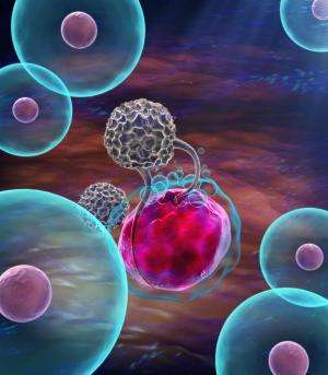
Mucins are glycoproteins that bear a high density of O-glycosylated serine and threonine residues. In species ranging from sea sponges to mammals, mucins are expressed at epithelial and endothelial surfaces where they defend against physical insults and pathogens1. The mechanisms by which mucins exert their functions at these surfaces fall broadly into two categories (Fig. 1a). First, mucins are critical to the initiation and propagation of biophysical signals. For example, their extended and rigid secondary structure enables their use by cells as force-sensitive antennae, as is the case for the mucin MUC1 during integrin-mediated adhesion to soft matrices2 and the mucin CD45 during macrophage pinocytosis3. Second, the glycopeptide epitopes presented by mucins act as ligands for various receptors, particularly those involved in cell adhesion and immune modulation4. For example, the immune cell receptor sialic acid-binding immunoglobulin-like lectin 7 (Siglec-7) binds the mucin CD43 on leukemia cell surfaces and delivers an immune inhibitory signal, analogous to classical checkpoint receptors such as PD-1 (ref. 5).
Cancers, especially carcinomas, hijack mucin signaling pathways to protect themselves from both biophysical and immunological insults. It is estimated that just one member of the mucin family, MUC1, is aberrantly expressed in more than half of carcinomas diagnosed per year in the United States6, a frequency matched by prototypical oncogenes such as RAS and MYC. In addition, common carcinomas, such as breast, ovarian and intestinal cancers, have mucinous forms, wherein tumor cells present as individual colonies suspended in a matrix of secreted mucin and polysaccharides7. As such, mucins have been exploited as tumor-enriched epitopes for antibodies8, antibody–drug conjugates9 and chimeric antigen receptor T cells10. In addition, antibodies, small molecules and peptides targeting the C-terminus of the MUC1 protein are under development to block its well-characterized cancer-driving functions, for example, by preventing dimerization11.
Generally, these mucin-targeted therapeutic interventions face the challenge that mucin signaling occurs through the cooperative action of hundreds of arrayed epitopes, a unique scaffolding secondary structure and a C-terminus capable of downstream growth and survival signaling. Therefore, it is attractive to consider strategies that enable therapeutic degradation of overexpressed mucins to reverse their pleiotropic tumor-progressive functions. Depletion of cellular mucins has hitherto only been achieved in the context of mucin 1 kidney disease, wherein a frameshifted and truncated form of MUC1 accumulates in early secretory compartments. This intracellular accumulation can be reversed with a small molecule that binds a cargo receptor, TMED9 (ref. 12).
Targeted protein degradation (TPD) has emerged as a powerful technique to address canonically undruggable targets. Classically, TPD uses bispecific molecules to traffic unwanted proteins to endogenous cellular proteolytic machinery for degradation. An advantage of this approach is that the aberrant protein is deleted, meaning that the full range of its pleiotropic effects on cell signaling are reversed13. As conventional TPD relies on proteasomal degradation, it is limited to targets that (1) can be bound with a bridging molecule that recruits endogenous degradation machinery and (2) contain cytosolic domains. Recently, cytosolic delivery of an exogenous, target-selective protease achieved proteolytic manipulation of cytosolic proteins without the need to recruit proteasome-shuttling pathways14, and leveraging of lysosomal-shuttling receptors has enabled TPD of cell-surface and secreted proteins15.
To degrade cancer-associated mucins, we developed a strategy for degradation of cell-surface proteins on specific cells without the need for endogenous degradation machinery. Specifically, a protease with dual glycan- and peptide-based selectivity for mucins is targeted to cancer cells via fusion to a camelid heavy chain variable domain (nanobody). We demonstrate that these targeted proteases reduce cancer cell viability in cellulo and blunt primary tumor burden and metastatic outgrowth in mouse breast cancer models. As nearly all extracellular proteins are glycosylated16 and glycosylation status is commonly altered in disease4, glycoform-dependent and cell-type-selective TPD presents a general opportunity for increasing on-target specificity for disease-driving extracellular proteins.
Results
Mucinase treatment undermines mucin-driven survival in cells
We and others have characterized proteases from the bacterial kingdom with selectivity for mucin domains17,18,19,20,21. These ‘mucinases’ act through recognition of joint peptide and glycan motifs, which have been mapped using mass spectrometry (MS) of cleavage products. As an initial candidate for therapeutic repurposing, we chose the zinc metalloprotease StcE from Escherichia coli serotype O157:H7. StcE recognizes the motif S/T*-X-S/T, where the first serine/threonine must bear an O-glycan (asterisk) for cleavage to occur18. StcE is agnostic to the structure of the glycan and the identity of the X amino acid, which can also be absent. StcE is therefore a pan-mucinase, which is able to act on epitopes present across the natural mucins.
To begin, we tested whether treatment with StcE could reduce cell viability by undermining the biophysical function of mucins. Expression of the MUC1 ectodomain in mammary epithelial cells induces a bulky glycocalyx, which causes the cells to lift from their basement membrane and thrive in suspension in a manner characteristic of circulating metastatic tumor cells22. To model suspension survival in cellulo, wild-type cells and cells overexpressing MUC1 were plated on ultralow-attachment plates treated with or without StcE and analyzed by flow cytometry to assess viability over 3 d (Fig. 1b). Under these anchorage-free conditions, StcE treatment resulted in rapid cell death, consistent with previously reported alterations in membrane biophysical signaling through PI3K–Akt (Fig. 1c)23. Meanwhile, StcE treatment of MUC1-expressing cells in standard tissue culture plates caused suspended cells to settle, after which they continued to divide, highlighting the low toxicity of mucinase treatment at nanomolar doses (Supplemental Videos 1 and 2).
Next, we asked whether StcE treatment could enhance immune surveillance of cancer cells. The mucin CD43 has been identified as a ligand on leukemia cells for the natural killer (NK) cell immune checkpoint receptor Siglec-7 (ref. 5). In this model, removal of CD43 potentiates NK cell killing of leukemia cell lines. To assess whether mucinase treatment would have a similar effect, we treated three leukemia cell lines with or without endotoxin-free StcE (Methods), incubated them with healthy human blood donor NK cells, and quantified viability after 4 h (Fig. 1d). StcE treatment resulted in loss of cell-surface CD43 and overall Siglec-7 ligand residency, as expected (Fig. 1e and Extended Data Fig. 1a,b)5. Demucinated leukemia cells were susceptible to increased NK cell surveillance, consistent with a recent report showing augmentation of breast cancer cell line killing by immortalized NK cells after mucinase treatment (Fig. 1f and Extended Data Fig. 1c,d)24.
As the presence of mucins on cell surfaces has been associated with decreased drug efficacy25, we also explored whether mucinase treatment would synergize with small-molecule cytotoxic drugs. Using scalable time-lapse analysis of cell death kinetics (STACK)26, we screened a 261-compound library in an ovarian cancer cell line (Extended Data Fig. 1e). The commercial library of 261 small molecules targeted a range of biological pathways (Supplemental Table 1) and had been used previously to evaluate treatments that modulate compound cytotoxicity27. Erastin, which induces ferroptosis through inhibition of the cystine:glutamate antiporter system xc–, scored among the top hits for compound cell death enhanced by StcE treatment (Extended Data Fig. 1f,g and Supplemental Table 1)28. A dose response with erastin2, a more potent analog, confirmed enhancement of ferroptosis with StcE cotreatment that was fully suppressed by the ferroptosis inhibitor ferrostatin-1. By contrast, there was no enhancement of ferroptosis induced by the mechanistically distinct compound RSL3 (Extended Data Fig. 1h)29. Taken together, these results demonstrate that removal of mucins via mucinase treatment can reverse their pleiotropic tumor-progressive roles.
Toxicity profile of StcE necessitates tumor targeting
Bacterial enzymes are currently used as frontline cancer therapeutics; for example, L-asparaginase from E. coli is employed in treatment of childhood acute lymphoblastic leukemias30. As mucinases had not, to our knowledge, been tested as injectable therapeutics, we assayed StcE for activity and tolerability in vivo. The maximum tolerated dose for StcE treatment in BALB/c and C57BL/6 mice was 0.25 mg per kg (body weight, unless indicated otherwise). Necropsy and complete blood count (CBC) analyses performed 3 h after injection of 15 mg per kg StcE revealed hemorrhages underneath the skull, ecchymoses throughout the gastrointestinal tract, neutrophil accumulation in the lungs and platelet depletion (Extended Data Fig. 2a and Supplemental Table 2). Western blotting using a mucin-specific probe19 showed that StcE injected at 0.25 mg per kg remained in circulation for at least 20 h and digested mucins throughout the body, though not as completely as at higher doses (Extended Data Fig. 2b–d). As endothelial and white blood cell surface mucins are critical components of clotting and immune activation pathways31, these findings established that an engineered mucinase variant with selectivity for tumor-associated mucins was necessary to avoid on-target, off-tumor effects.
The clinical success of antibody-drug conjugates has shown that fusion of toxic therapeutic cargo to antibodies is a viable strategy for lowering on-target, off-tumor toxicity and increasing on-target efficacy32. More recently, antibody-enzyme conjugates have been designed to target the hydrolytic activity of an enzyme to specific subsets of cells33. An important design principle of antibody-enzyme conjugates is to ensure that the activity of the enzyme is sufficiently low such that hydrolysis only occurs when the enzyme is concentrated at its target via binding of the antibody. Specifically, in previous work with an antibody of nanomolar affinity, micromolar enzymatic activity was shown to be effective for cell-surface targets34. As StcE is active at subnanomolar concentrations, our initial aim was to engineer a mucinase that retained its peptide and glycan specificity but exhibited activity within the micromolar range.
Structure-guided engineering reduces StcE activity and binding
We next used the previously published crystal structure35 and prior docking studies18 to rationally design a StcE mutant with reduced activity and cell-surface binding but retained specificity for mucins. To begin, we deleted two domains, the C and INS domains (Fig. 2a, left), as removal of the INS domain had previously been observed to reduce enzymatic activity, and removal of the C domain had been shown to decrease nonspecific cell-surface binding35, both of which were desirable for our targeted mucinase. We also mutated amino acids found near the active site that were not predicted to interact with key substrate residues in the docked complex. Specifically, W366, H367 and Y457 line the active site but do not directly interact with enzyme catalytic residues or substrate P2–P1′ residues, suggesting that mutation of these amino acids might reduce but not abrogate enzymatic activity (Fig. 2a, right, and Supplementary Fig. 1a)…


