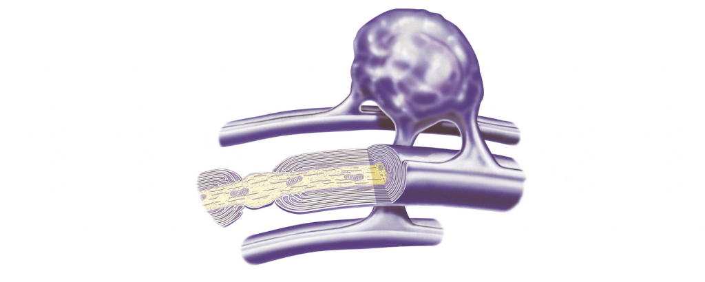
Highlights
- •Glia-derived chondroitin sulfate (CS) proteoglycans form aggregates of peri-synaptic matrix
- •Clusters of peri-synaptic matrix arise in an experience-dependent fashion
- •Versican core protein regulates the expression of a specific 6-sulfated CS glycoform
- •6-sulfated CS contributes to spatially organized synaptic refinement
Summary
Recent findings show that effective integration of novel information in the brain requires coordinated processes of homo- and heterosynaptic plasticity. In this work, we hypothesize that activity-dependent remodeling of the peri-synaptic extracellular matrix (ECM) contributes to these processes. We show that clusters of the peri-synaptic ECM, recognized by CS56 antibody, emerge in response to sensory stimuli, showing temporal and spatial coincidence with dendritic spine plasticity. Using CS56 co-immunoprecipitation of synaptosomal proteins, we identify several molecules involved in Ca2+ signaling, vesicle cycling, and AMPA-receptor exocytosis, thus suggesting a role in long-term potentiation (LTP). Finally, we show that, in the CA1 hippocampal region, the attenuation of CS56 glycoepitopes, through the depletion of versican as one of its main carriers, impairs LTP and object location memory in mice. These findings show that activity-dependent remodeling of the peri-synaptic ECM regulates the induction and consolidation of LTP, contributing to hippocampal-dependent memory.
Introduction
A critical challenge in neurobiology is to understand how neuronal activity refines local brain circuitry, resulting in experience-dependent learning. Converging evidence suggests that coordinated remodeling of adjacent synapses represents an efficient mechanism for the acquisition and storage of novel information.1,2,3,4,5,6 These studies show that the strength of a group of neighboring synapses on a dendritic segment increases within 90 min of a sensory stimulus.4,6,7 Thereafter, approximately 2 h following the triggering stimulus, the majority of newly potentiated synapses is reached by the protein product of the immediate-early gene (IEG) Arc/Arg3.1 (ARC), which drives massive activity-dependent heterosynaptic long-term depression (hLTD).4,5,7 As a consequence, only a small fraction of newly potentiated synapses are stabilized and functionally integrated into the local circuitry.4,5,6
This harmonized biphasic process results in an organized spatial arrangement of synaptic ensembles (i.e., synaptic clustering), putatively optimizing the signal-to-noise ratio within the local microcircuitry and providing a suitable substrate for the implementation of newly acquired information into the biological system.1,2,3,4,5,6
We reasoned that a coordinated remodeling of neighboring synapses requires massive restructuring of the local extracellular space, thus involving distinguishable changes in the peri-synaptic extracellular matrix (ECM). Indeed, converging findings show that specific ECM molecules, chondroitin sulfate proteoglycans (CSPGs), are key functional components of synaptic extracellular scaffolding, embedding pre- and post-synaptic terminals and powerfully regulating the actin cytoskeleton, synaptic plasticity,8,9,10,11,12,13,14,15,16,17 and memory consolidation.18,19 Accordingly,20 enzymatic digestion of CSPGs enhances the motility of dendritic spines,14 reinstates a juvenile-like form of experience-dependent plasticity,17 and promotes the erasure of fear memories.19 Importantly, previous studies showed that CSPGs’ role in synaptic plasticity largely depends on the chemical composition of glycosaminoglycan (GAG) chains, which can be alternatively sulfated in position 2, 4, or 6 of the N-acetyl-galactosamine sugar chains.10 Specifically, sulfation in position 6 was shown to positively mediate synaptogenesis, enhancing the establishment of synaptic contacts.11,21,22,23 This makes it an ideally suitable molecular player to participate in the dynamic and acute phase of local synaptic remodeling.
In primates and rodents, immunolabeling using CS56 antibodies recognizing 6- and 2,6-sulfated chondroitin sulfates24 results in morphologically conserved round structures populating cortical and subcortical brain regions,10,25,26,27 namely CS clusters (CSCs). While CSCs have been previously described in both human and rodent postmortem brain,25,26,27 their biological function remains unknown. Here, we show that, across species and brain areas, these structures present in distinct morphological conformations, from a cluster of sharply labeled converging processes (rosette CSCs; R-CSCs) to a group of diffuse puncta (diffuse CSCs, D-CSCs). We also show that the prevalence of R-CSCs and D-CSCs in the mouse barrel cortex (BCx) dynamically changes in response to naturalistic whisker stimulation, following a temporal profile consistent with the timing of synaptic structural plasticity. Consistently, we report that CSCs’ alternative conformations relate to specific geometrical features of dendritic spines and are paralleled by the dendritic expression of ARC. Finally, we discovered that the CS56 sulfation pattern is largely associated with the CSPG core protein versican (Vcan), and downregulation of Vcan selectively impairs the expression of chondroitin-6-O-sulfotransferase 2 (Chst7) in vitro and in vivo. Accordingly, hippocampal downregulation of Vcan–CS56 impairs long-term potentiation (LTP) and object location memory in adult mice, indicating that 6-sulfated CSPG Vcan plays a key role in learning, memory, and synaptic plasticity.
Results
CSCs show ultrastructural association with synapses and glial cells
We initially confirmed that immunolabeling using CS56 antibody on brain tissue resulted in widespread labeling of CSCs, which appeared along a spectrum of morphological configurations. For the purpose of this study, CSCs were classified into two main types, R-CSCs and D-CSCs, which correspond to the extremes of the whole morphological spectrum (Figure S1); this suggests that D-CSCs and R-CSCs may represent different transient stages of the same structure. Details for CSC morphological classification are reported in the STAR Methods (under “details for elements quantification in microscopy experiments,” subsection “CSCs classification”). Both D-CSCs and R-CSCs were detected in cortical and subcortical brain regions, across multiple mammalian species (human, non-human primates, and rodents; Figure S2). To investigate CS56 location in the synaptic scaffolding, we started by performing immunostaining with the CS56 antibody in the BCx, an area that is commonly densely populated by CSCs, in Thy1-eYFP mice,28 assessing their relationship with apical dendrites of layer 2/3. We found that CSCs encompass multiple dendrites (Figure 1A ) and that CS56-immunoreactive (CS56-IR) puncta are recurrently juxtaposed to Thy-1-labeled dendritic spines (Figure 1B). To further establish the broad-scale association between CS56 immunoreactivity and excitatory synapses, we quantified PSD95-IR post-synaptic elements juxtaposed to CS56-IR puncta within and outside of CSCs (Figure 1C). Within CSCs, 6% of PSD95-IR elements were contiguous to CS56-IR puncta. This percentage was significantly lower (3%) in equivalent, adjacent areas outside the CSCs (Figure 1D; Table S1). These findings indicate that CS56-reactive CSPGs form aggregates of peri-synaptic ECM and that this association occurs predominantly within CSCs.


