Highlights
- •ML infection of adult nine-banded armadillos promotes in vivo liver growth
- •Enlarged infected livers are functionally and architecturally normal without tumors
- •ML-induced partial reprogramming to progenitor/regeneration state drives liver growth
- •ML bypassing of normal liver growth restriction offers safer repair interventions
Summary
Ideal therapies for regenerative medicine or healthy aging require healthy organ growth and rejuvenation, but no organ-level approach is currently available. Using Mycobacterium leprae (ML) with natural partial cellular reprogramming capacity and its animal host nine-banded armadillos, we present an evolutionarily refined model of adult liver growth and regeneration. In infected armadillos, ML reprogram the entire liver and significantly increase total liver/body weight ratio by increasing healthy liver lobules, including hepatocyte proliferation and proportionate expansion of vasculature, and biliary systems. ML-infected livers are microarchitecturally and functionally normal without damage, fibrosis, or tumorigenesis. Bacteria-induced reprogramming reactivates liver progenitor/developmental/fetal genes and upregulates growth-, metabolism-, and anti-aging-associated markers with minimal change in senescence and tumorigenic genes, suggesting bacterial hijacking of homeostatic, regeneration pathways to promote de novo organogenesis. This may facilitate the unraveling of endogenous pathways that effectively and safely re-engage liver organ growth, with broad therapeutic implications including organ regeneration and rejuvenation.
Introduction
Adult organ growth promotion and rejuvenation are idealized strategies for treating dysfunction in disease, injury, or aging.1,2,3
Such strategies must engage highly coordinated multilineage functions in vivo. Although in vitro models, organoids, and mini-organs have potential for drug discovery, disease modeling, and regenerative medicine,4,5 they fail to model required organ-level complexity. Consequently, despite advances in such approaches,2,5 no current strategy achieves effective regrowth or rejuvenation of adult organs in chronic or aging-associated human diseases.
Liver is the exemplar organ for studying growth and regeneration.6,7
Unlike other solid organs, adult liver has the capacity to regain the prior mass after tissue loss, restoring homeostasis.7 In human chronic liver disease, repeated inflammatory injury and parenchymal cell death stimulate regenerative restitution of the liver cell mass in parallel with a wound-healing response.8
Some regenerative capacity remains in cirrhosis, although complete recovery is impossible and transplantation remains the only treatment. Chronic injuries are associated with increased risk for malignancy, which is highest in chronic viral infections.9,10
Endogenous pathways regenerating damaged liver remain poorly characterized, and failures of understanding contribute to a lack of pro-regenerative clinical strategies. As the health and economic burden of liver diseases rapidly increase,11,12 the absence of such repair strategies is critical. Moreover, the aging liver is more prone to progressive diseases as physiological functions decline.13,14,15
Maintenance of healthy liver for healthy aging is critical because it directly or indirectly influences other organ function, but there are no rejuvenating strategies slowing or reversing declining liver function during aging.
Current study of liver regeneration uses short-lived rodent models that require hepatocyte loss to stimulate regeneration8,16,17 that ceases when the original liver size is reached. Mechanisms stopping the response once the prior organ size is reached are unknown.17
The ability to bypass such upper-limit restriction would allow regeneration to be studied without prior liver injury. Understanding how regenerative machinery can be engaged de novo will provide paradigm-shifting adult organ regrowth and rejuvenation clinical strategies that could reduce or replace transplantation, but no such in vivo model is currently available.
Recent studies using the overexpression of the OSKM factors (Oct4, Sox2, Klf4, c-Myc) that originally generated induced pluripotent stem cells (iPSCs) from somatic cells18,19,20,21,22,23 showed a proof-of-principle that resetting committed cells to a progenitor stage of the same lineage permits tissue regeneration and rejuvenation. Therefore, alternative approaches that potentially increase adult tissue plasticity, proliferation, and de-differentiation should also be explored as strategies for tissue rejuvenation and regeneration.
Our studies on the biology of Mycobacterium leprae (ML)-host interaction24,25,26,27 led to the identification of ML’s natural ability to hijack the plasticity and regenerative properties of adult Schwann cells, partially reprogramming them into a progenitor cell/stem cell state beneficial to the bacteria.28
At the host level, ML-induced reprogramming promotes growth of infected tissues permitting bacterial propagation.24,25,28,29
These host-dependent features of ML without cytopathic or adverse effects during the establishment phase of infection permit use of ML as an evolutionarily adapted bacterial model for dissecting undefined host endogenous pathways.29,30, 31,32,33
Nine-banded armadillos (Dasypus novemcinctus) are New World placental mammals, the only mammal to produce four genetically identical/clonal litters and a natural host of ML.33,34,35
Experimental inoculation with viable ML produces disseminated infection,33,34,35,36,37 and their lifespan (12–13 years in the wild, up to 20 years in captivity) and core body temperature (32°C–35°C) are optimal for in vivo ML replication.33,34,35 Since their discovery as a natural host of ML,35 armadillos have been used for in vivo propagation of ML in the liver for harvesting bacteria for research.36
We explored whether this co-evolved bacterial pathogen in the liver of susceptible hosts exploited the same reprogramming strategies to expand host cells in vivo during natural infection as those observed in vitro in adult Schwann cells.28
We report a natural in vivo model of ML-infected nine-banded armadillos for mammalian adult liver growth at organ level without prior injury. We showed that bacteria-induced in vivo partial reprogramming significantly increased liver size with sustained function and architecture but without damage, fibrosis, or tumorigenesis during the establishment phase of infection. We define which cell types promote this organ growth and show that healthy liver lobule number, not size, with a proportionate expansion of the hepatocyte mass and vascular and bile ductal systems, is responsible. We delineate the molecular details to show evidence that ML have adapted dynamic partial reprogramming, regenerative, and developmental/fetal mechanisms to promote de novo liver organogenesis while maintaining tissue-protective and tumor-preventive strategies.
Results
In vivo ML infection of nine-banded armadillos promotes organ growth
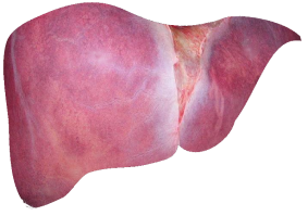
Adult nine-banded armadillo (>1.5–2 years old) livers with disseminated infection (“infected”) after injection of viable ML were compared with those from animals resistant to infection (“resistant”) and uninfected animals (Figures 1 and S1; Table S1; STAR Methods). During natural infection, ∼95% of humans and 20% of armadillos clear ML immediately, whereas infection progresses in the remainder. Clonal armadillo siblings (Figure 1A) were either fully resistant to infection or showed disseminated infection, indicating a strong heritable component for susceptibility and clearance. Resistant animals showed initial responses to infection, determined by serum ML-specific phenolic glycolipid-1 (PGL-1) antibody levels (Figure S1C; STAR Methods), but bacteria failed to propagate in the liver and only a few, presumably non-viable or dead, bacilli remained (Figure 1G; Table S1). Total liver/body weight ratio was significantly increased in armadillos infected for a period of 10–30 months compared with resistant (p < 0.0018) or uninfected animals (p < 0.001) (Figures 1B, 1D, and 1F). Livers from most infected animals showed a high bacterial count (up to 3.0E11 bacilli/g) (Figures 1C, 1F, 1G, and S1; Table S1). Liver/body weight ratio correlated with hepatic bacterial load in infected animals (Spearman’s rho [rs] = 0.5775764, p = 0.00007687; Figure 1E). Immunolabeling with an ML-specific anti-PGL-1 antibody and Wade-Fite acid-fast mycobacterial staining28,38 revealed ML in most hepatocytes and macrophages in small granulomas in infected animals (Figures 1C and S1D).


