Zoning in on liver growth
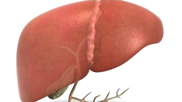
For organ homeostasis or regrowth after injury or disease, one or more stem cell populations is needed to rebuild lost tissue. There is considerable debate about the source of new cells in the liver. Two groups now identify the source of new hepatocytes (see the Perspective by Andersson). Although the liver may seem to lack major variation across its structure, its lobule is organized into concentric zones where hepatocytes express different metabolic enzymes. Wei et al. sought to systematically define the source of new liver cells by comparing 14 fate-mapping mice that label different liver cell types. They found that different regions of the liver lobule exhibit differences in hepatocyte turnover, with zone 2 representing a primary source of new hepatocytes during homeostasis and regeneration. Similarly, He et al. designed a genetic approach to record cell proliferation in vivo with high spatial and temporal resolution to enable continuous recording of proliferative events of any specific cell type at the whole-cell population level. Using this method, they identified zone 2 as having the highest proliferative activity and contributing the most to liver regrowth. These findings have implications for the cellular basis of chronic disease pathogenesis, cancer development, and regenerative medicine strategies.
Abstract
The liver is organized into zones in which hepatocytes express different metabolic enzymes. The cells most responsible for liver repopulation and regeneration remain undefined, because fate mapping has only been performed on a few hepatocyte subsets. Here, 14 murine fate-mapping strains were used to systematically compare distinct subsets of hepatocytes. During homeostasis, cells from both periportal zone 1 and pericentral zone 3 contracted in number, whereas cells from midlobular zone 2 expanded in number. Cells within zone 2, which are sheltered from common injuries, also contributed to regeneration after pericentral and periportal injuries. Repopulation from zone 2 was driven by the insulin-like growth factor binding protein 2–mechanistic target of rapamycin–cyclin D1 (IGFBP2-mTOR-CCND1) axis. Therefore, different regions of the lobule exhibit differences in their contribution to hepatocyte turnover, and zone 2 is an important source of new hepatocytes during homeostasis and regeneration.


