Abstract
In metastasis, cancer cells travel around the circulation to colonize distant sites. Due to the rarity of these events, the immediate fates of metastasizing tumor cells (mTCs) are poorly understood while the role of the endothelium as a dissemination interface remains elusive. Using a newly developed combinatorial mTC enrichment approach, we provide a transcriptional blueprint of the early colonization process. Following their arrest at the metastatic site, mTCs were found to either proliferate intravascularly or extravasate, thereby establishing metastatic latency. Endothelial-derived angiocrine Wnt factors drive this bifurcation, instructing mTCs to follow the extravasation–latency route. Surprisingly, mTC responsiveness towards niche-derived Wnt was established at the epigenetic level, which predetermined tumor cell behavior. Whereas hypomethylation enabled high Wnt activity leading to metastatic latency, methylated mTCs exhibited low activity and proliferated intravascularly. Collectively the data identify the predetermined methylation status of disseminated tumor cells as a key regulator of mTC behavior in the metastatic niche.
Main
Metastatic latency and tumor cell (TC) dormancy pose a major hurdle in the treatment of cancer1,2,3. During metastasis, latent TCs (LTCs) reside in close proximity to blood vessels and acquire a stem-like phenotype4. However, metastasizing tumor cells (mTCs) show remarkable heterogeneity in their genetic and molecular make-up, which can be attributed to certain cancer cells reaching a latent state whereas others outgrow immediately to form macrometastases5,6,7,8,9. This differential behavior is not solely driven by TC-intrinsic properties but is also influenced by this metastatic niche, because certain niches in principle favor TC proliferation whereas others are primarily tumor suppressive10,11,12. This argues for a scenario in which the induction of latency depends on cell-intrinsic properties and matching microenvironmental factors, which is also corroborated by the finding that disseminated TCs, once committed to a dormant fate, require dramatic events to be awakened13,14,15. We therefore hypothesized that TC behavior is primed during the initial arrival of mTCs at the metastatic niche and that the vascular endothelium, as the interface of dissemination, is a crucial fate instructor.
Wnt and epithelial-to-mesenchyme transition pathways drive extravasation and latency
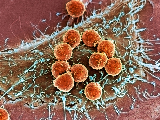
To test our hypothesis we developed an experimental model for temporal assessment of TC and endothelial cell (EC) interactions in the metastatic niche in vivo and at single-cell resolution. For this purpose, wild-type female BALB/c mice were intravenously injected with green fluorescent protein (GFP)-expressing 4T1 breast cancer cells (4T1-GFP). Lung-seeded TCs and corresponding total lung ECs were isolated at day 0 (baseline), day 1.5 (peak phase of TC extravasation) and day 3.5 (induction of TC proliferation) and enriched by fluorescence-activated cell sorting (FACS) (Fig. 1a and Extended Data Fig. 1a,b). Plate-based, single-cell RNA sequencing (scRNA-seq) was used to analyze the transcriptional signatures of TC–EC interactions during metastatic colonization. To discriminate extravascular from intravascular TCs, mice were intravenously injected at day 1.5 with fluorescently labeled anti-H-2Kd antibody. This antibody targets BALB/c-specific major histocompatibility complex I thereby labeling all body cells, including syngeneic 4T1-GFP cells that were exposed to the circulation, and hence creating a self-validating system. Cell types exposed to the circulation, such as circulating immune cells and ECs, showed positive staining in FACS whereas epithelial cells and tissue-resident immune cells remained unstained (Extended Data Fig. 1a). The approach was further validated using microscopy, which revealed that H-2Kd staining was confined to the vasculature and stained only TCs or parts thereof (for partially extravasated TCs) that were within the blood vessel (Extended Data Fig. 1c–e). Because TCs started to proliferate between days 3 and 4 postinjection (Extended Data Fig. 1b), they were further discriminated by their proliferation status at day 3.5 based on the dilution of CellTrace dye, independently of their extravasation status. TCs that retained CellTrace dye to the same extent as nonproliferative TCs from the day 1.5 time point were deemed latent whereas those with dye dilution, as evidenced by lower staining intensity in FACS, were considered proliferative (Fig. 1a and Extended Data Fig. 1b). Importantly, LTCs persisted in the lung for at least 2 weeks, indicative of a stable latent phenotype that resembled metastatic dormancy (Extended Data Fig. 1f). For each FACS-enriched TC subpopulation and matched ECs, equal cell numbers were sorted from at least three biological replicates. The cells in the dataset showed homogenous distribution for raw gene counts, normalized counts and detected genes (Extended Data Fig. 2a) and the replicates interlaced well in uniform manifold approximation and projection (UMAP) (Extended Data Fig. 2b,c), demonstrating the robustness of the experimental approach. Moreover, substructures in UMAP were found to be specifically enriched for cells from the respective TC FACS gates, suggesting that the gating strategy was suitable for enrichment of rare TC subpopulations even though it did not yield high purity (Extended Data Fig. 2b–f). Importantly, H2-K1 expression did not differ between the intravascular and extravascular fractions, highlighting that differences in staining intensity reflect exposure to circulation rather than gene regulation (Extended Data Fig. 2d).
Clustering of the combined TC dataset identified a total of five clusters enriched for cells of the respective FACS gates (Fig. 1b and Extended Data Fig. 2e,f). Trajectory analysis to reconstruct the colonization process with TCs of the intravascular cluster set as starting point identified three main trajectories, transitioning (1) from intravascular to proliferative cells, (2) to a subset of extravascular cells and (3) through a subset of extravascular cells to LTCs (Fig. 1c). This also reflected the pseudotemporal ordering of events, with the establishment of latency occurring last, suggesting that proliferation had preceded extravasation and latency induction (Extended Data Fig. 2g). Importantly, pseudotime correlated with real time, indicating that the trajectory analysis had faithfully recapitulated the sequence of biological events (Extended Data Fig. 2h–k). Moreover, extravascular TCs showed enrichment of genes that were previously shown to be involved in the extravasation process16 while LTCs were enriched for genes associated with cancer stemness and dormancy (Extended Data Fig. 2l,m and Supplementary Table 1). Taken together, these findings indicated that TC extravasation is a prerequisite for metastatic latency but dispensable for TC proliferation, which molecularly defines earlier microscopy-based concepts17,18,19. Importantly these data do not rule out extravascular TC growth, albeit strongly suggesting that intravascular cells are the major contributor to metastatic growth in the lung.
We next compared proliferative versus latent cells and scored each TC from the day 3.5 dataset for the expression of G2M- and S-phase genes. To exclude cells that had proliferated but dropped out of cycle, only TCs that showed dye retention and were not in cycle at the time point of sampling were considered as proliferation naïve and labeled as bona fide latent (Fig. 1d). Following regression of cell cycle-associated genes, differential gene expression analysis (DGEA) between bona fide latent and proliferative TCs was performed with subsequent gene set enrichment analysis (GSEA) (Fig. 1e and Supplementary Table 2). In line with previous reports20,21, transforming growth factor beta-signaling and epithelial-to-mesenchyme transition (EMT) gene sets were enriched in latent mTCs, suggesting early priming for tumor dormancy. Surprisingly, β-catenin-mediated canonical Wnt signaling was identified as one of the most significantly enriched pathways (Fig. 1e). Wnt ligands have been extensively characterized as protumorigenic growth factors22, promoting proliferation in both primary tumors and metastases as well as circulating TC (CTC) survival23,24,25,26. Unexpectedly, the expression of Wnt pathway- and EMT-associated genes was enriched alongside the extravascular–latency trajectory (Fig. 1f,g, Extended Data Fig. 2n,o and Supplementary Table 1), supporting the hypothesis that niche-derived Wnt ligands may drive the metastatic latency of a subset of mTCs that is characterized by a mesenchymal-like phenotype. Overall these data provide a transcriptional blueprint of mTC fate decisions in the metastatic lung.
Lung endothelium displays bimodal response following arrival of mTCs
To assess the endothelium’s response towards arriving mTCs, ECs of the combined dataset were first classified into known lung-specific EC subtypes—that is, general capillary ECs (gCaps), aerocytes (aCaps), cycling ECs and large-vessel ECs—based on previously reported cell marker gene expression27,28 (Extended Data Fig. 3a–i and Supplementary Table 1). For this purpose, each EC was scored for the expression of the marker gene sets and thresholds were set for classification of EC type (Extended Data Fig. 3f–i).
Next, DGEA of EC pseudobulks comparing individual experimental time points was performed, which revealed an immediate response pattern in gCaps with genes being mostly regulated at day 1.5 whereas aCaps showed little transcriptional dynamics (Fig. 2a–f and Supplementary Tables 3 and 4). To investigate the molecular basis of the observed gCap response, clustering analysis was performed, which revealed the emergence of a gCap subpopulation that clustered together with large-vessel ECs (Fig. 2e,f). DGEA between the emerging gCap cluster and gCap revealed upregulation of mostly metabolic and ribosomal genes, indicating enhanced biosynthesis and general activation (Supplementary Table 5). Significantly regulated genes were further filtered for temporal enrichment (increased on days 1.5 and 3.5 compared with day 0), and the gene expression profile of the resulting gene panel (Supplementary Table 1) was investigated in filtered capillary ECs (Fig. 2g,h). Interestingly, capillary ECs showed focal expression and upregulation of the gene panel, suggesting a subset of metabolically active ECs that produce biomass following TC arrival….


