In vitro embryo models supported by methods development in adjacent fields have revolutionized our understanding of embryogenesis.
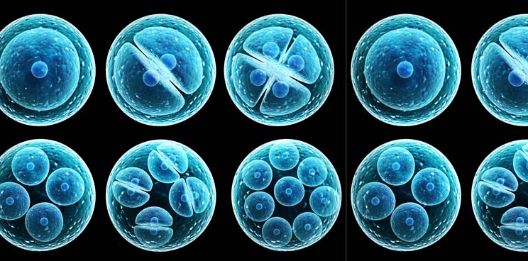
The process of development begins with a single cell that then progresses through multicellular forms and undergoes rounds of cell fate commitment and tissue patterning to ultimately form a complex body structure. The intricate mechanisms controlling the development of a zygote into a fetus during pregnancy have long been out of reach for researchers due to technical and ethical considerations. However, the last few years have seen tremendous advances in methods development that have shed light on these ‘black box’ events.
Pioneering methods in this space have successfully recapitulated key embryonic stages in vitro and thus reshaped our understanding of development. For example, two landmark papers1,2 in 2021 reported in vitro models of the human blastocyst, which is an early preimplantation developmental stage. Earlier this year a team reported3 a human perigastrulation model that recapitulates key events such as the formation of the amniotic and yolk sac cavities, early neurulation and organogenesis.
These models were further refined by incorporating extraembryonic tissues to study postimplantation events. For instance, two studies4,5 published in 2023 reported human stem cell-derived embryo models that could recapitulate embryonic events up to day 14 postimplantation in vivo.
These methods for modeling human development follows on the heels of technological advances and achievements first reported in mouse studies. Two seminal papers6,7 described in vitro models derived from mouse stem cells that could recapitulate natural mouse embryos in utero up to 8.5 days postfertilization. These embryos developed until the organogenesis stage, co-developing with extraembryonic tissues. The complexity of this model, which mimicked natural embryos in morphological and transcriptomic analysis, led to more authentic organ and tissue development. However, while conclusions can be drawn from studying developmental processes in animal models, species specific features of human embryonic development can be easily missed in such cases.
For the remarkable insights into embryogenesis enabled by these sophisticated models, we have chosen methods for modeling development as our Method of the Year 2023.
In this special issue, a Comment8 by Magdalena Zernicka-Goetz provides an in-depth look at the latest embryo models that have been reported. These models are poised to help researchers to investigate the molecular mechanisms behind morphogenesis and the signaling cues that underlie the tissue patterning. Zernicka-Goetz warns researchers that while these embryo models lead to novel insights, they are far from perfect recapitulations of the in vivo embryo. Indeed, it is imperative to be aware of the limitations of a system and how it might affect the inferences that are drawn.
The fidelity of an in vitro model must always be verified against in vivo-derived tissue. In the case of embryonic tissue, this poses a particular challenge due to the limited availability of embryos for research as well as ethical issues surrounding human embryo manipulations. A Comment9 by Muzlifah Haniffa and colleagues discusses how the advent of single-cell multiomic technologies and the subsequent developmental cell atlases have served as essential benchmarks for testing the validity of an embryo model.
Beyond the development of cell atlases, single-cell technologies have also led to the emergence of sophisticated methods for lineage tracing and trajectory analysis. Although these methods have not yet been extensively used for human embryonic models, they have been instrumental in mapping cell fate events, such as in human brain organoids10 and zebrafish embryos11. In a Comment12, Bushra Raj describes recent methodological advances in this field and its potential for studying snapshots of development.
The state-of-the-art embryo models have been bolstered by decades of research into methods for the in vitro culture of mammalian embryos. Hongmei Wang and colleagues explore this in their Comment13. They write that to optimize culture conditions that support an in vitro embryo, there is still a need for the development of biomaterials that mimic the physiological microenvironment of the embryos, as well methods to study the mechanical environment experienced by cells during the stages of embryogenesis.
This opinion is echoed by Idse Heemskerk and colleagues in another Comment14, which discusses recent methods that now allow researchers to map the forces within a developing embryo. The Comment explores how embryo models have the potential to shed light on the interplay between tissue mechanics, patterning and morphogenesis. Related to this, a research paper in this same issue from Hervé Turlier and colleagues reports foambryo15, a method for performing force inference from 3D fluorescence images of mouse and ascidian embryos. A second research paper, by Noah Mitchell and Dillon Cislo, presents TubULAR16, a tissue cartography method for analyzing deformations in dynamic tissues during processes such as morphogenesis.
A nascent approach to studying embryonic development is computational embryology. Researchers explore how to computationally generate and perturb virtual embryos that are built from experimental data. A research article17 from the team of Patrick Müller describes a deep learning-based approach to grade the similarities between embryos at different time points. In an earlier issue of Nature Methods, the same team published EmbryoNet18, a neural network that can analyze zebrafish embryo phenotypes and link them to major signaling pathways. Our News Feature19 also checks in with researchers in this area, who share hopes and challenges concerning digital embryos and their future.
In a discussion about advances in embryo research, it would be remiss to not consider the ethical implications of engineering human embryo models. In their Comment20, ethicists Nienke de Graeff, Lien De Proost and Megan Munsie explain the new ethical questions posed by embryo models. They discuss whether embryo models should be held to the same guidelines as organoids or whether their developmental potential affords them a new status.
Embryo models, especially human systems, are a new and exciting frontier supported by parallel methods development in the fields of single-cell omics, biomaterials and mechanobiology. We must have an open and transparent dialog about the limitations of these methods as well as the ethical risks of in vitro human embryos models. Meanwhile, we hope you are with us as we recognize the potential these methods have in not only unraveling the details of embryogenesis but also modeling development and pregnancy-related disorders.


