Abstract
In the early stages of mitosis, cohesin is released from chromosome arms but not from centromeres. The protection of centromeric cohesin by SGO1 maintains the sister chromatid cohesion that resists the pulling forces of microtubules until all chromosomes are attached in a bipolar manner to the mitotic spindle. Here we present the X-ray crystal structure of a segment of human SGO1 bound to a conserved surface of the cohesin complex. SGO1 binds to a composite interface formed by the SA2 and SCC1RAD21 subunits of cohesin. SGO1 shares this binding interface with CTCF, indicating that these distinct chromosomal regulators control cohesin through a universal principle. This interaction is essential for the localization of SGO1 to centromeres and protects centromeric cohesin against WAPL-mediated cohesin release. SGO1–cohesin binding is maintained until the formation of microtubule–kinetochore attachments and is required for faithful chromosome segregation and the maintenance of a stable karyotype.
Main
During mitosis, the duplicated genome needs to be accurately distributed over the two daughter cells. The cohesin protein complex holds together the sister DNAs from replication until mitosis1,2,3. Cohesin entraps DNA inside its ring-shaped structure4, which at its core consists of SMC1, SMC3 and SCC1 (also known as RAD21 or Mcd1). SCC1 is bound by either of two paralogous HEAT repeat-containing proteins, SA1 or SA2 (also known as STAG1 and STAG2)5.
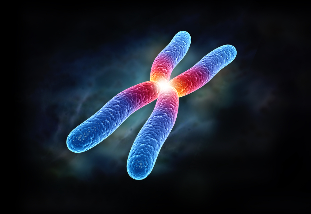
Cohesin complexes have a dynamic mode of DNA binding that involves DNA entrapment and release. From DNA replication until mitosis, the cohesin complexes that hold together the sister DNAs are locked on DNA to render cohesin resistant to cohesin’s release factor WAPL6. During mitosis, cohesin is removed from chromosomes in two waves. First, cohesin is removed from chromosome arms in a WAPL-dependent manner through a process known as the prophase pathway7,8,9,10. Cohesion at centromeres is protected by Shugoshin (SGO1)11,12,13,14, giving rise to the typical X-shaped structure of human chromosomes. SGO1 protects centromeric cohesin by recruiting PP2A to counteract cohesin phosphorylation by mitotic kinases, and SGO1 also directly competes with WAPL for cohesin binding15,16,17,18. Centromeric cohesion is maintained until proper attachment of microtubules to the kinetochores, upon which the remaining cohesin is cleaved by separase to trigger anaphase onset19. By protecting centromeric cohesion, SGO1 thus ensures faithful chromosome segregation.
Cohesin has a dual role, as it not only holds together sister DNAs but also builds the DNA loops that shape the interphase genome. To control this latter function, cohesin is bound by the architectural factor CTCF20. We showed recently that CTCF binds to cohesin through a conserved YxF motif in the amino terminus of CTCF. This CTCF segment interacts directly with a composite binding interface formed by the SA2 and SCC1 subunits of cohesin21. The SA2 interface is conserved from fungi to mammals and is known as the conserved essential surface (CES)22. For simplicity, we refer to the composite SA2–SCC1 binding pocket as the CES. The direct interaction of the YxF motif of CTCF with the CES is required for formation of CTCF-anchored loops at TAD boundaries21. It has also been suggested that the CES region interacts directly with both SGO1 and WAPL18. The interaction of SGO1 with cohesin is promoted by phosphorylation of SGO1 T346. However, this phosphorylation does not control the direct interaction between the CES and SGO1 (refs. 17,18). SGO1 contains a YxF motif close to this phosphorylation site that could be key to this interaction. In agreement, previous biochemical experiments have shown that an SGO1 fragment containing the YxF motif can directly compete with CTCF for binding to the CES21. If so, cohesin complexes may be controlled through a shared mechanism, irrespective of whether these complexes build DNA loops or rather hold together the sister DNAs.
We report here the X-ray crystal structure and AlphaFold model of the cohesin subcomplex SA2–SCC1 bound to a fragment of SGO1 (Fig. 1a). We demonstrate that SGO1 engages the CES of SA2–SCC1 through its YxF motif. The binding mode is similar to that seen with the YxF motif from CTCF. Mutations that abolish SGO1–CES interaction interfere with the localization of SGO1 to centromeres and lead to severe cohesion defects. We infer that engagement of the CES surface of cohesin by distinct chromosomal regulators is a universal principle that allows control of cohesin function during different chromosomal processes.
Results
Structural basis for SGO1–cohesin interaction
Previous data indicate that SGO1 interacts directly with the SA2–SCC1 subunit of cohesin18. The interacting region contains a YxF motif that is conserved in vertebrate SGO1 proteins (Fig. 1b and Extended Data Fig. 1a). Phosphorylation of T346, probably by CDK1 (ref. 17), enhances the interaction with SA2–SCC1 but is not essential for binding18. We were able to obtain crystals with a SGO1 peptide spanning amino acids 331–341 containing the YxF motif but not with an extended peptide spanning amino acids 331–349 containing pT346. We determined the X-ray structure by molecular replacement to a minimum Bragg spacing of 3.2 Å (Table 1). An Fo–Fc omit electron-density Fourier map exhibited clear features of the SGO1 peptide (Extended Data Fig. 1b). The SGO1 peptide is bound to the CES binding pocket (Fig. 1c and Extended Data Fig. 1c–e). Amino acid residues F337 and Y335 of SGO1 bind into hydrophobic pockets using a similar binding mode to that seen previously for CTCF21. Briefly, the binding pocket for F337 of SGO1 contains amino acids S334, I337 and L341 from SCC1 and Y297 and W334 from SA2 (Fig. 1c and Extended Data Fig. 1d). Y335 of SGO1 binds in a deep hydrophobic pocket containing L329, L366 and F367 (Fig. 1c and Extended Data Fig. 1e). A model calculated using AlphaFold structure prediction23 showed an almost identical binding mode and suggested additional interactions between SGO1 amino acids 341–349 and SA2 (Fig. 1c).
Accordingly, isothermal titration calorimetry (ITC) experiments showed that the T346-phosphorylated SGO1 fragment spanning amino acids 331–349 bound SA2–SCC1 with a lower equilibrium dissociation constant (2.3 ± 0.4 μM) compared with a nonphosphorylated SGO1 peptide spanning amino acids 331–341 (13.5 μM ± 1.4) (Fig. 1d). Using glutathione S-transferase (GST) pulldown experiments, we found that SGO1 retained SA2–SCC1 on GST beads (Fig. 1e). Mutation Y335A or F337A of SGO1 abolished the interaction. Mutation of critical CES amino acid residues including SA2 W334A, R370Q, SCC1 I337A L341A or the absence of SCC1 also impaired SGO1 binding. Together, our data confirm the previous biochemical mapping of SGO1 interaction18. We conclude that the YxF motif of SGO1 is essential for binding to the composite CES of SA2–SCC1. Phosphorylation of SGO1 at T346 enhances the interaction.
The SGO1–CES interaction protects centromeric cohesion
To test whether the SGO1–cohesin interaction that we identified in our crystal structure controls sister chromatid cohesion, we mutated the endogenous SGO1 allele in HAP1 cells using CRISPR–Cas9 technology. We thereby obtained HAP1 cells with SGO1Y335A F337A as their sole copy of SGO1 (Extended Data Fig. 2a–d). We then analyzed sister chromatid cohesion in these cells by performing chromosome spreads. Wild-type cells, as expected, displayed robust sister chromatid cohesion. SGO1Y335A F337A cells, however, displayed severe cohesion defects (Fig. 2c,d). Correspondingly, a large proportion of these cells failed to form a proper metaphase plate, leading either to mitotic slippage or mitotic catastrophe (Extended Data Fig. 3a,b)….


