Abstract
Current gene therapy for Duchenne muscular dystrophy (DMD) utilizes adeno-associated virus (AAV) to deliver micro-dystrophin (µDys), which does not provide full protection for striated muscles as it lacks many important functional domains of full-length (FL) dystrophin. Here we develop a triple vector system to deliver FL-dystrophin into skeletal and cardiac muscles. We split FL-dystrophin into three fragments linked to two orthogonal pairs of split intein, allowing efficient assembly of FL-dystrophin. The three fragments packaged in myotropic AAV (MyoAAV4A) restore FL-dystrophin expression in both skeletal and cardiac muscles in male mdx4cv mice. Dystrophin-glycoprotein complex components are also restored at the sarcolemma of dystrophic muscles. MyoAAV4A-delivered FL-dystrophin significantly improves muscle histopathology, contractility, and overall strength comparable to µDys, but unlike µDys, it also restores defective cavin 4 localization and associated signaling in mdx4cv heart. Therefore, our data support the feasibility of a mutation-independent FL-dystrophin gene therapy for DMD, warranting further clinical development.
Introduction
Duchenne muscular dystrophy (DMD) is a fatal genetic disease afflicting approximately 1 in 3800–6300 live male births and caused by genetic mutations in the DMD gene located on the X chromosome1. The dystrophin protein encoded by the DMD gene belongs to the spectrin superfamily of cytoskeletal proteins2. The major isoform expressed in striated muscles is a 427 kDa protein, containing four main domains: an N-terminal actin-binding domain, a long rod-like domain composed of spectrin-like repeats, a cysteine-rich domain that binds to dystroglycan and other membrane-associated proteins, and a C-terminal domain that interacts with syntrophin and α-dystrobrevin, forming a large dystrophin-glycoprotein complex (DGC)3,4. The DGC links the actin cytoskeleton to the extracellular matrix, facilitating force transmission, providing mechanical stability and membrane protection, and mediating signal transduction5,6,7. Disrupted expression of dystrophin leads to repeated cycles of muscle damage and repair, which eventually exhaust the regenerative capacity of muscle stem cells and cause fibrosis and fatty replacement. The progressive loss of muscle mass and function in the skeletal, cardiac, and respiratory muscles eventually leads to impaired mobility, cardiomyopathy, respiratory failure, and early death8,9.
A gene replacement therapy using adeno-associated virus (AAV) to deliver a correct copy of the DMD gene could allow the restoration of dystrophin to halt/reverse the deterioration of muscles10. However, the limited cargo capacity of AAV (~4.5 kb) makes it challenging to deliver the FL DMD gene (cDNA is over 11 kb). Early studies found that patients carrying a large internal deletion of the DMD gene developed a milder form of the disease, known as Becker muscular dystrophy (BMD), due to the expression of truncated yet partially functional dystrophin proteins11. These findings led to the development of micro-dystrophins (μDys) gene therapy, which aims to restore the expression of a truncated version of the dystrophin protein12,13,14. These miniaturized forms of dystrophin are only about 1/3 of the FL-dystrophin, retain some of the essential functional domains of the protein and can be packaged into one AAV vector for in vivo delivery. This approach has shown promise in preclinical animal models, restoring muscle integrity and improving muscle function13,14,15. Recently, the US Food and Drug Administration (FDA) granted accelerated approval to Sarepta’s Elevidys (μDys delivered in AAVrh.74) for DMD patients aged 4 to 5 years16. However, due to the lack of 2/3 of the dystrophin coding sequence containing critical rod and hinge domains of dystrophin needed to connect with other proteins in the dystrophin complex, these μDys gene therapies are unable to provide full protection of skeletal and heart muscle integrity and function15,17,18,19. This limitation highlights the urgent need to develop novel strategies to deliver larger or even FL-dystrophin.
Several different approaches have been explored using dual AAV vectors to deliver larger payloads, including fragmented genome assembly, overlapping, trans-splicing, and hybrid approaches. For instance, two vectors containing an overlapping fragment to express large transgenes may undergo homologous recombination20,21,22,23. Upon co-delivery, the efficiency of homologous recombination is, however, a limiting factor for this strategy. Another approach known as trans-splicing is to take advantage of the natural concatamerization ability of AAV to expand the transgene size using dual AAV vectors24,25. However, the head-to-tail concatamer formation and the trans-splicing of the pre-mRNA across the ITR junction pose rate-limiting steps. These approaches may be combined as shown for hybrid vector systems26. Despite the varying success of these dual AAV vector strategies in preclinical studies, the efficiency of expression from the available dual-vector systems is insufficient for many clinical gene therapy applications. It is even more challenging to deliver the FL-dystrophin sequence, which is about three times the packaging capacity of AAV, thus requiring at least three vectors to package the entire coding sequence. A previous attempt with triple trans-splicing AAV vectors showed the feasibility of expressing FL-dystrophin, albeit with only very low efficiency27.
Split inteins are small polypeptides that self-assemble and undergo a protein trans-splicing (PTS) reaction, resulting in the formation of a mature, fully functional protein in a “traceless manner”. We and others have successfully delivered the oversized base editors in dual AAV vectors in vivo using the split intein strategy28,29,30. Leveraged on this success, we developed a triple AAV system with orthogonal split inteins to deliver FL-dystrophin protein. Packaged into an engineered myotropic AAV capsid31 (MyoAAV4A), this triple vector combination restored the expression of FL-dystrophin and significantly improved muscle histopathology and function in a mouse model of DMD.
Results
Proof-of-concept design of split intein constructs to assemble FL-dystrophin
The cDNA for FL-dystrophin is over 11 kb, about three times of the AAV packaging capacity. Thus, three AAV vectors are required to package the entire coding sequence of FL-dystrophin. We rationally split FL-dystrophin cDNA into three fragments based on (1) the fragment size, (2) the protein domain structure, and (3) the junctional sequence compatibility for split intein. We previously utilized Cfa32 and Gp41-133, two orthogonal inteins with a remarkably fast rate of protein trans-splicing (PTS), to successfully assemble adenine base editor (ABE)28. We, therefore, chose these two orthogonal pairs of inteins for our initial test. The first split site was chosen in between spectrin repeat (SR) 8 and 9, where a cysteine residue is followed by a bulky tryptophan residue as required for efficient protein splicing by the Cfa intein (Fig. 1a). The second split site was selected within the end of Hinge 3 (H3) domain, where two consecutive serine residues are located, which may facilitate protein splicing by the Gp41-1 intein (Fig. 1a). In addition, this is also where the native Dp140 isoform starts. We named the three fragment constructs of this first version as Dys-N1, Dys-M1, and Dys-C1, respectively. All expression cassettes utilized a mini-CMV promoter with a muscle creatine kinase enhancer (meCMV). Transfection of Dys-N1/M1/C1 into HEK293 cells resulted in the expression of FL-dystrophin detectable by three different anti-dystrophin antibodies that specifically recognize the N, M, or C fragments, respectively (Fig. 1b). However, the FL-dystrophin band was much weaker than the unassembled or partially assembled dystrophin fragments. Varying the ratio of the three plasmids improved the relative abundance of the FL versus the unassembled or partially assembled dystrophin signals, with the 4:2:1 ratio of N1:M1:C1 plasmids yielding the highest level of FL-dystrophin. The C-terminal fragment band was much more intense than either the FL or MC bands, indicating that the assembly between the M and C fragments mediated by Gp41-1 intein needs to be improved.
Optimization of split intein constructs to improve FL-dystrophin assembly
Encouraged by this initial observation, we attempted to optimize the assembly between the M and C fragments. First, we tested a different split-site (IGA-SPT) within the H3 domain and mutated the −1 position from alanine to tyrosine to favor the Gp41-1-mediated PTS (Fig. 2a). These changes (Dys-M2 and Dys-C2) led to a substantial improvement in the FL-dystrophin assembly (Fig. 2b, c) with reduced unassembled (Fig. 2d–f) and partially assembled fragments (Supplementary Fig. 1a–c). In addition, we removed a small intron within the C fragment construct (Dys-C3), which significantly increased the FL-dystrophin band intensity (Fig. 2b, c). Next, we reasoned that the +1 to +3 position on the extein (e.g., Dys-C3) may also affect the PTS efficiency. We thus mutated the SPT sequence to SSS at the junction site on the C fragment construct (Fig. 2g). In addition, we changed the strong Kozak sequence to a weak one considering the abundance of the C fragment. However, these two changes (Dys-C4) led to a substantial reduction of the FL-dystrophin band with an increased accumulation of the NM band and C band (Fig. 2h–l and Supplementary Fig. 1d–f). We reasoned that this might be caused by the use of the weak Kozak sequence, which potentially leads to the translation initiation from a downstream in-frame start codon so that the C fragment was expressed without the intein fusion. Indeed, after we switched the weak Kozac sequence back to a strong one in this version of the C fragment construct (Dys-C5), FL-dystrophin expression was restored (Fig. 2h–l and Supplementary Fig. 1d–f). Of note, we also added a synthetic signal for ubiquitin-dependent proteolysis (PB29)34 in the Dys-C5 construct to lower the expression level of the C fragment. Although the FL-dystrophin band signal was reduced when compared to Dys-C3, we observed that the NM fragment was almost undetectable, suggesting that these changes improved NM and C assembly. We further tested a different intein (IMPDH−1), which has a fast PTS rate comparable to Gp41-1 with a native junction sequence of GGG-SIC33, similar to the split site of dystrophin (IGA-SPT) (Fig. 2m). We also mutated alanine at the -1 position of Dys-M2 extein to glycine to further mimic the native junction sequence of IMPDH-1. These changes (Dys-M3 and Dys-C6) significantly improved the FL-dystrophin signal by ~86% (Fig. 2n, o). Moreover, the unassembled C fragment band was dramatically reduced by ~76% as compared to Dys-N1/M2/C5 (Fig. 2n, r), with marginal effects on the unassembled N and M fragments (Fig. 2n, p, q) and partially assembled fragments (Supplementary Fig. 1g–i). Finally, we inserted two different poly-adenylation signal sequences35 at the upstream of the start codon in order to further lower the expression level of the C fragment (Dys-C7 and Dys-C8) (Supplementary Fig. 2a). These changes significantly reduced the C-fragment band intensity while still maintaining a high level of FL-dystrophin expression (Supplementary Fig. 2b, c, f) and similar N/FL (Supplementary Fig. 2d) or MC/FL ratio (Supplementary Fig. 2i). However, we found that lowering C-fragment expression further caused a concomitant accumulation of the M (Supplementary Fig. 2e) and NM (Supplementary Fig. 2g, h) fragments. These data suggest that some excess of the C-fragment favors the assembly of FL-dystrophin.
In contrast to the canonical inteins, atypical inteins with small inteinN and large inteinC have also been discovered36,37. To test whether the atypical inteins could also enable the assembly of FL-dystrophin, we modified the N, M, and C constructs using the atypical inteins Cat38 and VidaL39 to mediate the N-M and M-C splicing, respectively (Supplementary Fig. 3a). Western blotting analysis showed that these atypical inteins can also confer the assembly of FL-dystrophin, but the efficiency was much lower than Dys-N1/M3/C6 (Supplementary Fig. 3b). We chose the best-optimized version Dys-N1/M3/C6 for further in vivo studies.
Restoration of FL-dystrophin expression in mdx 4cv mice following systemic MyoAAV delivery of Dys-N1/M3/C6
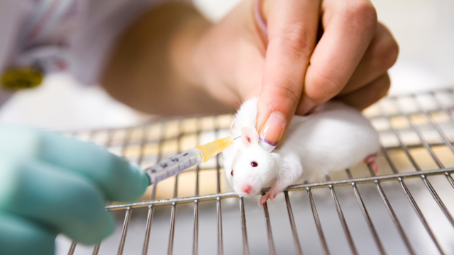
We first replaced the generic promoter meCMV with a synthetic muscle-specific promoter Spc5-1240 (for the N and M fragment construct) or Spc2-2640 (for the C fragment construct) for AAV packaging. We chose a recently engineered myotropic AAV capsid, MyoAAV4A, for packaging because of its superior muscle and heart transduction in mice and monkeys following systemic delivery31. A total dose of 2E + 14 vg/kg AAV vectors consisting of N1, M3, and C6 at a 2:1:1 molar ratio was delivered into a cohort of mdx4cv mice (N = 13) at the age of 3–4 weeks via retro-orbital injection (Fig. 3a).
Immunofluorescence staining was performed using the aforementioned anti-dystrophin antibodies (Fig. 3b). Dystrophin expression could be detected at the sarcolemma of wild-type (WT) gastrocnemius (GA) muscle with all these three antibodies, while GA muscle from mdx4cv mice was negative for any of these antibody stains (Fig. 3b). AAV administration restored dystrophin expression that can be detected by all these three antibodies (Fig. 3b). Importantly, dystrophin signals were correctly localized at the sarcolemma without noticeable accumulation in the cytoplasm. On average, dystrophin was detected in 83.4 ± 2.7% muscle fibers (Fig. 3c). Dystrophin expression was also robustly rescued in the cardiac muscles of mdx4cv mice with 78.3 ± 2.7% cardiomyocytes being dystrophin positive following AAV administration (Supplementary Fig. 4a, c). However, dystrophin+ fibers in diaphragm muscles (8.6 ± 2.6%) were much lower than those in the GA muscles (Supplementary Fig. 4b, d).
Western blotting was also performed to substantiate these observations. Again, FL-dystrophin was readily detectable using the three different N-, M-, or C-recognizing antibodies in GA muscles from mdx4cv mice treated with AAV-N1/M3/C6 (Fig. 3d). Interestingly, both the N- and M-recognizing antibodies detected mostly the FL-dystrophin with weak partially assembled dystrophin fragments, while the unassembled N- or M-fragment was hardly discernable (Fig. 3d). The C-recognizing antibody detected FL-dystrophin and unassembled C-fragment with roughly equal intensities, while the partially assembled MC fragment was almost undetectable (Fig. 3d). FL-dystrophin expression was also readily detectable in the heart muscles of AAV-treated mdx4cv mice (Supplementary Fig. 4e), but the diaphragm muscle showed a much weaker expression of FL-dystrophin and the C fragment following AAV treatment (Supplementary Fig. 4f), likely reflecting weak activity of the Spc2-26 promoter and/or MyoAAV4A capsid in diaphragm muscle.
The loss of dystrophin in dystrophic muscle severely affects the integrity of the entire DGC5,6,7. To test if AAV-N1/M3/C6 treatment restores the other components of the DGC in mdx4cv muscles, we performed immunofluorescence staining with the antibodies against various components of the DGC such as α-sarcoglycan (α-SG), β-SG, α-dystroglycan (α-DG), β-DG, neuronal nitric oxide synthase (nNOS), and α-dystrobrevin (α-DB). As shown in Fig. 3e, the DGC components, including α-SG, β-SG, α-DG, β-DG, nNOS, and α-DB, were all severely reduced at the sarcolemma of GA muscle fibers from mdx4cv mice but were substantially restored by AAV-N1/M3/C6 treatment.
Functional and histopathological improvement in mdx 4cv mice following systemic MyoAAV4A delivery of Dys-N1/M3/C6
Increased muscle injury and reduced muscle force production are the pathological hallmarks of DMD. To examine if the AAV-N1/M3/C6 treatment improves the muscle pathologies in mdx4cv mice, we first measured the serum creatine kinase (CK) levels at 5 weeks following AAV administration. As compared to WT mice, mdx4cv animals showed a dramatic elevation in serum CK (WT: 220.6 ± 109.1, n = 14 vs mdx4cv: 3946.0 ± 341.1, n = 12; p < 0.0001, Fig. 4a), which was significantly reduced in AAV-treated group (1060.0 ± 229.4, n = 13; p < 0.0001), suggesting that FL-dystrophin expression reduces muscle injury in dystrophic mice. To test if AAV-N1/M3/C6 treatment improves muscle function, we measured the muscle contractility using an in vivo muscle test system28,41,42. The maximum plantarflexion tetanic torque was measured during supramaximal electric stimulation of the tibial nerve at 150 Hz. As previously shown, mdx4cv mice produced greatly reduced torque as compared to WT controls (WT: 544.7 ± 9.8, n = 8 vs mdx4cv: 298.2 ± 10.6, n = 10; p < 0.0001, Fig. 4b). Systemic delivery of AAV-N1/M3/C6 significantly increased the tetanic torque in mdx4cv mice by ~51.7% (452.5 ± 12.0, n = 11; p < 0.0001, Fig. 4b)….

