Immune cells called macrophages commonly function as scavenger-like (phagocytic) cells that ingest and remove damaged cells. Writing in Nature, Culemann et al.1 report that the macrophages present in joints also fulfil an unexpectedly different role.
Macrophages derive from two main cellular lineages2. One lineage arises from bone-marrow-derived immune cells called monocytes. The other lineage is monocyte independent, and is derived from cells that disperse into the tissues during embryonic development2. The tissue-resident macrophages in this lineage have distinctive gene-expression profiles3,4 that depend on the particular tissue in which they reside.
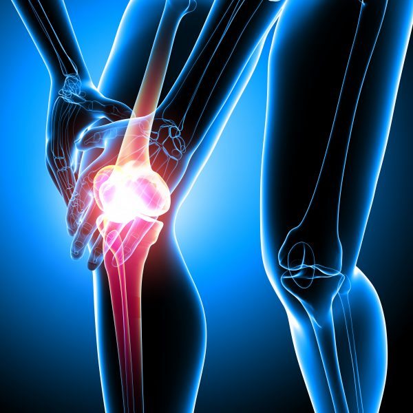
Rheumatoid arthritis is an immune-mediated disease associated with inflammation and the destruction of the cartilage and bone in joints, and macrophages have a key role in the initiation of this condition. However, little is known about the relative contribution of the two lineages of macrophages to the development and function of joints in health and disease. To add to the complexity, macrophages exist as various subsets, some of which are pro-inflammatory, whereas others are anti-inflammatory and aid tissue repair5.
To study macrophages, the authors began by focusing on a protein called CX3CR1, which is expressed on monocytes and macrophages. The authors engineered CX3CR1-expressing cells in mice to make a red fluorescent protein so that the cells could be tracked in vivo. These cells were monitored in knee joints using an approach called 3D light-sheet fluorescence microscopy, and the joint tissue was treated using a technique that enabled the authors to obtain ‘optical clearance’, which improves the visualization of internal structures6.
Unexpectedly, the authors’ observations revealed that CX3CR1-expressing macrophages exist as a layer of cells that forms a barrier, similar to a thin protective membrane, in the healthy joint (Fig. 1). This barrier forms as an outer layer of cells in the synovium, a region of the tissue that lines the joint. The barrier layer forms in a part of the synovium called the lining layer, and it physically separates the synovial fluid (which bathes the joint) from the sublining layers of the synovium. The CX3CR1-expressing barrier-forming macrophages are found adjacent to a layer of cells called fibroblasts in the lining layer….


