Highlights
- •Age and APOE4 enrich for TIM, microglia co-expressing stress and inflammatory markers
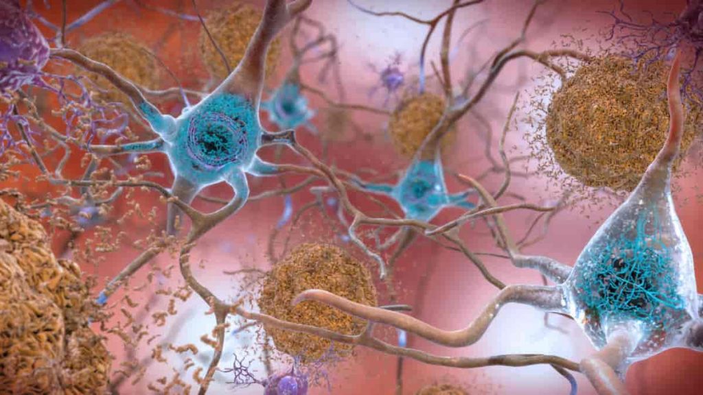
- •TIM are present in human AD and spatially colocalize with Aβ plaques in the cortex
- •TIM are an exhausted-like population with impaired function and signaling
- •Aducanumab treatment alters TIM heterogeneity and state in an APOE-dependent manner
Summary
The dominant risk factors for late-onset Alzheimer’s disease (AD) are advanced age and the APOE4 genetic variant. To examine how these factors alter neuroimmune function, we generated an integrative, longitudinal single-cell atlas of brain immune cells in AD model mice bearing the three common human APOE alleles. Transcriptomic and chromatin accessibility analyses identified a reactive microglial population defined by the concomitant expression of inflammatory signals and cell-intrinsic stress markers whose frequency increased with age and APOE4 burden. An analogous population was detectable in the brains of human AD patients, including in the cortical tissue, using multiplexed spatial transcriptomics. This population, which we designate as terminally inflammatory microglia (TIM), exhibited defects in amyloid-β clearance and altered cell-cell communication during aducanumab treatment. TIM may represent an exhausted-like state for inflammatory microglia in the AD milieu that contributes to AD risk and pathology in APOE4 carriers and the elderly, thus presenting a potential therapeutic target for AD.
Introduction
Alzheimer’s disease (AD) is an incurable neurodegenerative disease and the predominant form of dementia, characterized by progressive synaptic dysfunction, neuronal loss, and cognitive decline. Excessive accumulation of the amyloid-β (Aβ) peptide and aggregates of hyperphosphorylated tau proteins are the major pathological features of the Alzheimer’s brain, followed by neuroinflammation, which is thought to be driven primarily by microglial cells.
Microglia are yolk-sac-derived myeloid cells and the dominant immune population of the brain.4,5,6 Although microglia are central mediators of classical neuroinflammation, they are also a highly heterogeneous population, existing in the AD brain in distinct states that can be differentially beneficial or detrimental to disease progression.7 Nonetheless, the factors determining whether a given microglial population either constrains or contributes to AD pathology remain poorly defined.
Apolipoprotein E (APOE) is a secreted protein named for its central role in lipid trafficking with three common human isoforms: APOE2, APOE3, and APOE4. Although these alleles only vary at two amino acid sites, they are nonetheless strongly associated with differential risk for several diseases, including hyperlipidemia and atherosclerosis.15 APOE is of particular interest in human health due to its role as the single largest monoallelic risk factor for late-onset AD (LOAD), with APOE4 increasing risk and APOE2 reducing risk relative to APOE3, the most common allele in the population.16 APOE is also a regulator of immunity more broadly, with roles in anti-tumor immunity,17,18 the response to SARS-CoV-2 infection,19 and multiple other inflammatory contexts.20 Still, the mechanisms by which brain immune cells are jointly modulated by aging and distinct APOE alleles are not well understood.
We aimed to characterize the immune cellular changes in the AD brain driven by aging and distinct APOE alleles by generating a single-cell atlas of immune cells from the brains of AD mice bearing APOE2, APOE3, or APOE4 alleles at distinct ages. Our atlas encompasses mice from 10 weeks of age to ∼2 years of age, an understudied super-elderly state. By combining two global genome-wide modalities across multiple time points in AD progression, we systematically profiled the complex dynamics of the neuroimmune system and leveraged these data to interrogate its emergent biological properties. We identified a population of microglia expressing a signature of inflammatory and stress signaling markers whose frequency was enriched by age and APOE4 genotype. These microglia are present in human AD; exhibit impaired capacity for Aβ clearance; and appear to be key participants in the microglial response to aducanumab, an approved AD treatment. These findings identify a putative exhausted-like state for microglia in AD and a potential target cell for future therapeutic intervention.
Results
Single-cell RNA sequencing identifies a TIM state in AD mice
We crossed 5×FAD mice, a common murine model of AD progression, with mice bearing human APOE2, APOE3, or APOE4 alleles in the murine Apoe locus (hereafter denoted as AD∗APOE2, AD∗APOE3, and AD∗APOE4). We flow-sorted Cd45+ cells from the hippocampal and cortical regions of these APOE-homozygote 5×FADhet mice at 10 and 20 weeks of age, as well as of APOE3– and APOE4-homozygote 5×FADhet mice aged to 96 weeks (∼2 years, equivalent to a human octogenarian), pooling cells from 3 to 6 mice per group. We then performed 10X single-cell RNA sequencing (scRNA-seq) using either 3′v3 or 5′v2 capture technology, yielding an atlas of 30,868 single cells after filtering and quality control (Figure 1A). We sorted microglia specifically from hippocampal and cortical tissues due to their central roles in AD pathology and progression.3,21,22,23 Reciprocal principal component analysis mediated integration24 followed by clustering identified 17 clusters, including one major cluster representing ∼80% of single-cell profiles that expressed microglial markers8 such as P2ry12 and Tmem119 (Figures 1B and S1A). Subclustering this microglial group generated 20 discrete microglial subpopulations, including clusters corresponding to the disease-associated microglia (DAM) state25 that were identified by high expression of genes such as APOE and Cst7 (Figure 1C). We augmented these 20 clusters by leveraging a k-nearest neighbors (KNN) approach26 to survey the single-cell manifold for small, highly connected groups of cells with unique expression signatures. After hierarchical statistical evaluation, a single high-confidence microcluster of 11 cells expressing a transcriptional program including interleukin (Il)-34 and Ano1 passed thresholding (Figures S1B and S1C). We manually annotated this cluster as Il-34+ microglia, arriving at a final set of 21 clusters. To establish that the clusters identified in this pipeline are meaningful and not spurious products of overclustering, we trained a 100-ply random forest classifier on a 4-fold cross-validation scheme over 25 iterations on the raw count data from each cluster, using the classifier to produce a pairwise confusion matrix between each combination of microglial subclusters. Cells were rarely misassigned by this classifier (Figure S1D), supporting the final clustering generated by this approach.


