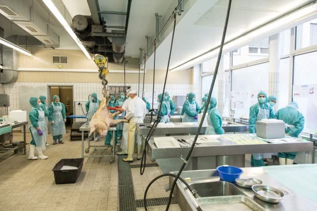
First out is a kidney: its dark red fades to beige as it is washed of its blood. The pancreas, harder to find amid the tangle of inner organs, is rushed on to dry ice. Speed is essential because tissues degrade after death and each detail counts in this autopsy.
The precious organs belong to Boar 1339, which, for 3.5 years, had lived a normal pig’s life on the farm of a German university, despite the diabetes it was born with. Earlier this month, the animal was killed, and the body parts placed in the service of science, as part of a growing movement to maximize the scientific benefits of every animal used in research.
Thousands of tiny tissue and fluid samples from the boar’s 226-kilogram body now sit in the newly constructed Munich MIDY-PIG Biobank in Germany the world’s first systematic repository of tissue from a large, genetically engineered, non-human animal.
The biobank, part of Munich’s Ludwig Maximilian University (LMU), houses tissue taken from throughout the body and from pigs of different ages. It is designed to help diabetes researchers to discover the molecules and mechanisms involved in the long-term complications of the disease, including the degeneration of small blood vessels and nerves, heart and kidney disease, and blindness. These develop over a lifetime, and are poorly understood.
“We are more similar to pigs than we like to think, so this resource will be very valuable,” says diabetes researcher Patrik Rorsman at the University of Oxford, UK. The samples are freely available to researchers anywhere in the world, who need only pay for the postage.
As pressure grows to reduce the number of animals used in research, biobanks are becoming attractive because they allow teams working on different organs and aspects of the disease to use the same animal. “Biobanking means that no part of the animal is wasted,” says LMU geneticist and veterinary surgeon Eckhard Wolf, who launched the pig biobank.
Only a few animal biobanks have so far been built, and most are for mice. Pigs, although more expensive to house and breed, could be more useful because of their larger size and the greater similarity of their physiology and metabolism to those of humans.
To create the pig biobank, scientists used genetic engineering and cloning techniques to create animals with a damaged gene called MIDY, which means that they need a daily insulin injection. The animals were then bred with healthy pigs so that, on average, half of the second-generation litters had diabetes and the other half were healthy, and thus able to serve as experimental controls.
The 12th addition to the bank from this process, Boar 1339 was already anaesthetized when it arrived in the cavernous autopsy room of the 101-year-old veterinary school in a leafy suburb of Munich. Waiting for the pig was a team of 25 veterinary surgeons and technicians, gowned up, masked and alert at parallel dissection tables.
A hoist raised the pig by its hind legs and pathologists injected the animal with a lethal dose of anaesthetic. Then, in swift, precision choreography, the team moved in with their knives, cleavers, hammers and pincers. They removed organs, muscles, nerves and more, and transferred the tissues to the tables at which the waiting dissectors set about their complicated sampling process, strictly following an 80-page protocol. “We don’t want to complete a super-sophisticated sampling and then realize we forgot to weigh the liver,” says Andreas Blutke, one of the chief pathologists at the LMU. The chopping, hacking and puncturing are loud; the concentration of the workers keeps the background noise to a murmur. The whole process takes a mere 2 hours 15 minutes.
Because the cells in any organ are of different sizes, shapes and orientations, and are unevenly distributed, the team takes numerous samples using different methods to ensure that the whole organ is appropriately represented. The system is so sophisticated that it gives researchers a three-dimensional anatomical reconstruction of the exact cell types.
Crucially, the researchers divide each sample and preserve the portions in different ways, each optimized for either structural or molecular analysis. This allows both types of analysis to be done on the same sample. “I think this is the only service which allows you to do molecular profiling and cellular anatomy from the same sample,” says Wolf.
As soon as Rorsman heard about the biobank, he saw its benefit. He suspects that the long-term complications of diabetes are caused by changes in a particular molecule in the cells of several different tissues. Material from the biobank will allow him to confirm that he is on the right track, he says, before he obtains samples from human biobanks, which takes a long time because of ethical constraints. Herbert Tempfer, a diabetes researcher at the University of Salzburg in Austria, is already analysing samples of tendons notoriously fragile in people with diabetes from the biobank.
Others want to know how similar pig diabetes is to the human disease. Immunologist Åsa Hidmark at the University of Heidelberg in Germany made the three-hour journey to Boar 1339’s dissection to cut samples of skin from its trotter. She hopes to discover that the nerve endings in the outer layer have been lost, as happens in people with diabetes.
The ultimate value of the bank will depend on how much it is used and there are no guarantees. Despite its collection of 42 tissues taken from 940 mouse lines, a mouse biobank at the Wellcome Trust Sanger Institute near Cambridge, UK, has so far received only 50 or so requests for material. “There is a lack of awareness of its value,” says Jacqui White, who leads this Sanger mouse-autopsy project.
Wolf plans to extend his biobank to other genetic pig models as they are developed. Next in line are probably pigs engineered to have Duchenne muscular dystrophy. A sow implanted with a cloned genetically modified embryo is now pregnant.


