Some cancer cells can survive chemotherapy by resorting to cannibalism.
By “consuming” neighboring cancer cells, some cells have found a way to obtain the energy they need to remain alive and induce relapse after a course of chemotherapy is completed, according to new findings published in the Journal of Cell Biology,
Breast cancer cells and doxorubicin
Doxorubicin (the chemotherapy drug used in this study) is as an anthracycline. This type of chemotherapy drug works by damaging the DNA in a cancer cell, achieved via three different mechanisms:
By binding to DNA via intercalation between base pairs on the DNA helix
By preventing repair of DNA by inhibiting an enzyme called topoisomerase II
By acting as a powerful iron-chelator. This property enables the formation of iron-doxorubicin complexes that can bind both the cell’s DNA and its membrane
Crafty cancer cells
However, some cells are able to survive initial treatment with doxorubicin, which can result in relapsed tumors. These “crafty” cells transform into senescent cells, entering a “dormant” state, whilst remaining metabolically active. Senescent cells can encourage the growth of a tumor by releasing tumor-promoting factors and inflammatory molecules.
This is particularly problematic when treating breast cancer cells that retain a “normal” copy of the TP53 gene. TP53 is mutated in just 30% of breast cancers – the remaining 70% with “normal” TP53 can escape death in response to chemotherapy-induced DNA damage. Breast cancer survival can be predicted by TP53 mutation status – those with TP53 wild-type tumors have poorer survival.
“Understanding the properties of these senescent cancer cells that allow their survival after chemotherapy treatment is extremely important,” explains first author Crystal A. Tonnessen-Murray, in a recent press release.
Study design and findings
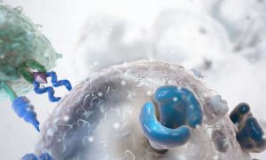
In the new study the team discovered that the breast cancer cells exposed to doxorubicin (or other chemotherapy drugs) which then transformed into senescent cells often engulfed their neighboring cancer cells.
“We used cells and tumors that express fluorescent proteins (e.g., GFP) of different colors to examine interactions between cells in treated tumors and also in treated cultures,” explains James G. Jackson, corresponding study author.
Jackson elaborates on some of the techniques used in the study: “We used confocal microscopy, which allows one to visualize a 3-dimensional representation of an image. This allowed us to determine if cells were under, on top of, or actually within another cell. We also used time lapse microscopy to generate movies of cells cannibalizing.”…


