Years after Elizabeth Blackburn, Ph.D. won the Nobel Prize for her seminal work on the molecular nature of telomeres and co-discovery of the ribonucleoprotein enzyme telomerase, scientists still struggle with fully understanding the intricacies surrounding this inherent genetic mechanism for maintaining genomic stability. The telomerase enzyme plays an important role in aging and most cancers, but until recently many aspects of the enzyme’s structure could not be resolved.
Now, scientists at UCLA and UC Berkeley have generated high-resolution images of telomerase, providing major insight into the enzyme’s functionality and possibly leading toward new directions for treating cancer and preventing premature aging.
“Many details we could only guess at before, we can now see unambiguously, and we now have an understanding of where the different components of telomerase interact,” explained senior author Juli Feigon, Ph.D., professor of chemistry and biochemistry at UCLA. “If telomerase were a cat, before we could see its general outline and the location of the limbs, but now we can see the eyes, the whiskers, the tail, and the toes.”
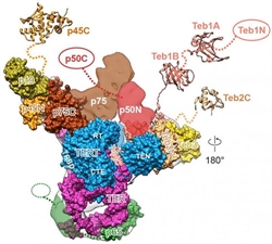
“Structure of Tetrahymena telomerase reveals previously unknown subunits, functions, and interactions.”
Telomeres the ends of eukaryotic chromosomes that serve as protective caps vital for preserving the genetic information maintain their genomic integrity through the activity of telomerase. When the enzyme is not active, telomeres shorten after each round of replication, eventually leading to cell death.
Conversely, cells with abnormally active telomerase can constantly rebuild their protective chromosomal caps and become immortal a state that at first blush seems appealing, but is, however, detrimental to the cell due to the accumulation of DNA errors over time. This is often the exact scenario for many cancers, allowing them to grow and spread uncontrollably.
When Dr. Feigon and her laboratory initially set their sights on telomerase, they merely wanted to learn how telomerase works. Fighting cancer or slowing the aging process were not areas they had considered delving into.
“Our research may make those things achievable, even though they were not our goals,” Dr. Feigon remarked. “You never know where basic research will go. When telomerase and telomeres were discovered, no one had any idea what the impact of that research would be. The question was, ‘How are the ends of our chromosomes maintained?’ We knew there had to be some activity in the cell that does that.”
Dr. Feigon and her team filled in the pieces of the telomerase puzzle using the ciliated protozoanTetrahymena thermophila, which they found has much more analogous telomerase enzyme to humans than previously thought.
“This is the first time that a whole telomerase directly isolated from its natural workplace has been visualized at a sub-nanometer resolution, and all components are identified in the structure,” stated co-lead author Jiansen Jiang, Ph.D., a UCLA postdoctoral scholar.
Previously, researchers had thought that telomerase was comprised of eight sub-units: seven proteins and RNA. But Dr. Feigon and her colleagues discovered two additional proteins, Teb2 and Teb3 that increase telomerase’s activity.
Additionally, the investigators found that within the enzyme’s catalytic core, which is formed by the RNA and its partner proteins TERT and p65, the RNA forms a ring around the donut-shaped TERT protein. Moreover, the scientists observed that a key protein called p50 interacts with several components of telomerase, including TERT, Teb1, and p75 all having important implications for telomerase’s function.
The UCLA and UC Berkeley teams are continuing to push forward and fill in even more details of the telomerase puzzle with the hope that their research could lead to the development of new pharmaceuticals that target specific subunits of telomerase structure.
“There is so much potential for treating disease if we understand deeply how telomerase works,” Dr. Feigon concluded.


