Highlights
- •The N terminus of ApoE binds to PIPs and TAG in an isoform-specific manner
- •Lean astrocytes secrete cholesterol-loaded ApoE; this is unaffected by polymorphism
- •Fatty astrocytes chronically exposed to fatty acids produce TAG-rich ApoE particles
- •APOE4 boosts TAG secretion, whereas APOE2 is less active and behaves as APOE-KO
Summary
Apolipoprotein E transports lipids and couples metabolism between astrocytes and neurons. The E4 variant (APOE4) affects these functions and represents a genetic predisposition for Alzheimer’s disease, but the molecular mechanisms remain elusive. We show that ApoE produces different types of lipoproteins via distinct lipidation pathways. ApoE forms high-density lipoprotein (HDL)-like, cholesterol-rich particles via the ATP-binding cassette transporter 1 (ABCA1), a mechanism largely unaffected by ApoE polymorphism. Alternatively, ectopic accumulation of fat in astrocytes, a stress-associated condition, redirects ApoE toward the assembly and secretion of triacylglycerol-rich lipoproteins, a process boosted by the APOE4 variant. We demonstrate in vitro that ApoE can detect triacylglycerol in membranes and spontaneously assemble lipoprotein particles (10–20 nm) rich in unsaturated triacylglycerol, and that APOE4 has remarkable properties behaving as a strong triacylglycerol binder. We propose that fatty APOE4 astrocytes have reduced ability to clear toxic fatty acids from the extracellular milieu, because APOE4 reroutes them back to secretion.
Introduction
The strongest genetic risk factor for late-onset Alzheimer’s disease is a polymorphism in the major brain lipid transporter, apolipoprotein E (ApoE), and one of its putative receptors, triggering receptor expressed on myeloid cells 2 (TREM2) (Corder et al., 1993; Guerreiro et al., 2013; Jonsson et al., 2013). ApoE is a lipid-binding protein that transports lipids between different organs and/or cell types. In the periphery, ApoE is an essential component of the principal lipoproteins: chylomicrons, very low-density lipoproteins (VLDLs), and high-density lipoproteins (HDLs) (Getz and Reardon, 2009; Mahley and Rall, 2000). In the brain, ApoE is produced locally at sites of stress or injury, such as excitotoxicity (Xu et al., 2006), but the lipoprotein particles involved in ApoE function, and more generally in lipid homeostasis, remain poorly characterized. ApoE exists as three common isoforms in humans. They differ at amino acid positions 112 and 158: APOE2 has two cysteines, APOE3 has a cysteine at position 112 and an arginine at 158, whereas APOE4 has arginines at both positions and represents the strongest genetic risk factor for late-onset, sporadic Alzheimer’s disease (Corder et al., 1993). Early work showed that, in the blood, APOE4 preferentially associates with VLDL, whereas APOE3 displays a preference for HDL (Gregg et al., 1986; Steinmetz et al., 1989; Weisgraber, 1990).In the brain, ApoE contributes to a bidirectional metabolic network that couples astrocytes and neurons (Ioannou et al., 2019; Liu et al., 2017; Qi et al., 2021). Astrocytes release ApoE in the form of HDL-like particles that transport cholesterol, phospholipids, hydrophobic vitamins, and antioxidants to neurons. This ApoE-mediated flux of hydrophobic molecules supports neuronal activity, remodeling, and repair (Bu, 2009; Huang et al., 2004). ApoE is also an essential component of a recently discovered detoxification mechanism that couples astrocytes and neurons. Hyperactive neurons secrete excess fatty acids (FAs) and peroxidized FAs (toxic by-products of their metabolism) in the form of ApoE lipid-containing particles that are taken over by astrocytes (Ioannou et al., 2019). In astrocytes, toxic FAs are stored in lipid droplets and metabolized in the mitochondria by β-oxidation.A consensus has emerged that APOE4 represents a hypomorphic variant that is less effective in detoxifying and feeding the neurons. Astrocytes expressing APOE4 have an increased number of lipid droplets (Farmer et al., 2019; Sienski et al., 2021), diminished cholesterol secretion, and impaired neurotrophic functions (Rawat et al., 2019; Yamazaki et al., 2019; Zhao et al., 2017). APOE4 is also less effective than the other isoforms in maintaining the coordinated detoxification pathway between neurons and astrocytes (Qi et al., 2021). However, the underlying molecular mechanisms remain elusive. Here, we have studied the molecular mechanisms of ApoE lipidation in astrocytes, the main producers of this protein in the brain, and evaluated the influence of ApoE genetic polymorphisms associated with Alzheimer’s disease to the lipidation process. We show that, in astrocytes, ApoE mainly forms HDL-like particles, rich in cholesterol and phosphatidylcholine, via a pathway involving the ATP-binding cassette transporter A1 (ABCA1). We also observed dramatic changes in ApoE lipidation once astrocytes accumulated excess fat, a disturbance often seen in aging brains or following stress. Under those conditions, ApoE formed lipoprotein particles, rich in unsaturated triacylglycerol, and, remarkably, APOE4 proved to be much more effective for this process. Overall, our data support a new model in which the apparent loss of detoxifying and feeding function of APOE4 could actually result from its high aptitude to assemble potentially toxic triacylglycerol-rich particles.
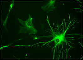
Results
In vitro, ApoE binds phosphoinositides and triacylglycerol in an isoform-specific manner
ApoE is known to bind PI(4,5)P2 -containing membranes in a process related to its lipidation by the ABCA1, but the molecular mechanisms and the effect of polymorphism remain unclear (Chernick et al., 2018; Gulshan et al., 2016; Krimbou et al., 2004; Wahrle et al., 2004). To better understand the effects of ApoE polymorphism on its lipidation, we measured its ability to recognize a range of specific membranes and lipids in vitro, in the absence of a functional cellular machinery. For this, we used a microfluidic device coupled to automated fluorescence microscopy (Saliba et al., 2014). ApoE contains two independently folded domains. The carboxy terminus contains a lipoprotein-binding site (Nguyen et al., 2009), whereas the amino terminus, which is made of several amphipathic, helical structures, contains the LDL-receptor-binding region (Croy et al., 2004). These helical structures are often present in apolipoproteins and known to interact with specific membranes or lipids (Gulshan et al., 2016). Therefore, truncated versions of ApoE that contained only the amino-terminal helical structures and that carry the two polymorphic sites were analyzed (N-APOE2, N-APOE3, and N-APOE4). We purified the recombinant, sfGFP-tagged versions of APOE2, APOE3, and APOE4 and of their respective amino termini from Escherichia coli (Figure S1A). The thermal stability of the different ApoE variants—measured by nano differential scanning fluorimetry (nanoDSF)—indicates that they were all properly folded (Figure S1B). Discrete differences in the thermal stability of the three isoforms—with APOE4 being the least thermostable—were also observed and reflect distinct structural properties (Acharya et al., 2002).We measured the recruitment of ApoE variants to different types of simple, artificial membranes made of a carrier lipid—phosphatidylcholine (the most abundant lipid in eukaryotic cells)—and increasing amounts of specific signaling lipids (Figures 1A , 1B, and S1C). The analysis was systematic, comprising the major lipid classes. It consisted of over 1,000 individual protein-lipid-binding experiments. Interestingly, none of the ApoE variants tested recognized cholesterol embedded in membranes (Figure S1C). However, consistent with previous reports (Gulshan et al., 2016), we observed an efficient recruitment of ApoE to membranes in the presence of phosphoinositides (PIPs), especially PI(4,5)P2 and PI(3,4,5)P3 (Figures 1A and 1B). The effect of PIPs was specific, as the binding did not occur when other negatively charged lipids, such as phosphatidylserine, phosphatidic acid, or sphingosine-1-phosphate, were present (Figure S1C)….


