Abstract
There is a growing appreciation that the direct interaction between bacteriophages and the mammalian host can facilitate diverse and unexplored symbioses. Yet the impact these bacteriophages may have on mammalian cellular and immunological processes is poorly understood. Here, we applied highly purified phage T4, free from bacterial by-products and endotoxins to mammalian cells and analyzed the cellular responses using luciferase reporter and antibody microarray assays. Phage preparations were applied in vitro to either A549 lung epithelial cells, MDCK-I kidney cells, or primary mouse bone marrow derived macrophages with the phage-free supernatant serving as a comparative control. Highly purified T4 phages were rapidly internalized by mammalian cells and accumulated within macropinosomes but did not activate the inflammatory DNA response TLR9 or cGAS-STING pathways. Following 8 hours of incubation with T4 phage, whole cell lysates were analyzed via antibody microarray that detected expression and phosphorylation levels of human signaling proteins. T4 phage application led to the activation of AKT-dependent pathways, resulting in an increase in cell metabolism, survival, and actin reorganization, the last being critical for macropinocytosis and potentially regulating a positive feedback loop to drive further phage internalization. T4 phages additionally down-regulated CDK1 and its downstream effectors, leading to an inhibition of cell cycle progression and an increase in cellular growth through a prolonged G1 phase. These interactions demonstrate that highly purified T4 phages do not activate DNA-mediated inflammatory pathways but do trigger protein phosphorylation cascades that promote cellular growth and survival. We conclude that mammalian cells are internalizing bacteriophages as a resource to promote cellular growth and metabolism.
Introduction
Bacteriophages, also called phages, are viruses that infect and kill bacteria, their natural hosts. Phages are ubiquitous across environments and are intrinsic components of our microbiomes, colonizing all niches of the body [1]. As such, the human body is frequently and continuously exposed to a diverse community of phages [2,3]. This is especially true within the gut, which houses a high-diversity microbial community [4]. Phages are essential components of the gut and participate in the genetic diversification and individualization of the gut microbiome throughout our life span [5–10]. While phages facilitate many changes to these gut microbial communities, they are also known to interact with the underlying mammalian cell layers [1,3,11–13]. Mammalian cells can engulf phages via a variety of mechanisms, leading to the internalization and accumulation of active phages [3,12–14]. Phages have been shown to bind specific mammalian cellular receptors, triggering receptor-mediated endocytosis [15,16]. However, the predominant mechanism by which phages have been shown to enter mammalian cells is through nonspecific internalization via macropinocytosis [1,12,13].
Macropinocytosis is an actin-based process characterized by the nonspecific internalization of extracellular fluid, nutrients, and potential microorganisms in large endocytic vesicles known as macropinosomes. Macropinocytosis is essential for cellular growth and cell proliferation as it allows the cell to access extracellular nutrients [17–21]. Additionally, macropinocytosis can sample and subsequently detect pathogens and foreign nucleic acids, leading to the activation of the innate immune system [22]. Macropinocytosis of phages by mammalian cells is a nonspecific process whereby cells create actin-mediated ruffles elongating outwards from the cytosol to engulf the extracellular milieu and any phages residing within it. Phages internalized via this pathway steadily accumulate within intracellular macropinosomes [13]. The downstream processing of the macropinosome can follow various pathways, including fusion with other endocytic vesicles, fusion with lysosomes leading to acidification and the inactivation of internalized components, recycling and transport to plasma membranes, and constitutive exocytosis [20].
Once inside the cell, phages may stimulate a diverse array of effects. The few studies that have investigated the cellular and innate immune response to phages have hinted at 2 opposing responses. On the one hand, certain phages like T4 or T7 are known to induce anti-inflammatory responses [23–27]. On the other, a growing number of studies have shown pro-inflammatory immune responses and inflammation to specific phages [28–30], including the filamentous phage Pf [29]. As such, it appears that certain phages may induce anti- or pro-inflammatory responses, highlighting an underlying specificity with the cellular detection of specific viral types. It remains mechanistically unclear how phages interact with and modulate the mammalian cells’ innate immune response and how these interactions can influence downstream cellular processes.
In this study, we investigate whether phage T4 can modulate cellular and innate immune pathways across in vitro cell lines. We demonstrate that phages were internalized by mammalian cells via macropinocytosis, with functional phages continually accumulating within macropinosomes [12,13]. All phage preparations were highly purified and confirmed to be free of bacterial endotoxins [31,32]. To further ensure the cellular responses detected were elicited by the phages themselves, we used a comparative control that consisted of the highly purified phage lysate filtered 4 times through a 0.02-μm filter to remove phage particles and obtain a phage-free lysate composed of the background supernatant. Using these samples, we performed luciferase assays, cellular proliferation assays, and interrogated antibody microarrays to probe the cellular and innate immune changes induced by the presence of T4 phage. From these results, we showed that T4 phage do not activate DNA-mediated inflammatory pathways, and rather, that phage internalization induced broad cellular signaling cascades affecting cell metabolism, survival, cell cycle progression, and growth.
Results
T4 phage do not activate the intracellular DNA-sensing receptors TLR9 and cGAS-STING
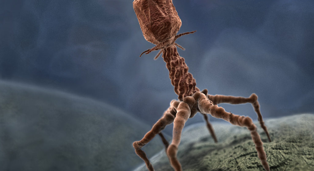
We focused on bacteriophage T4, a virulent Tevenvirinae phage, with an approximately 200-nm long myovirus morphology and a 168,903-bp genome, which infects Escherichia coli [33,34]. This phage was selected as it was previously demonstrated to be internalized by mammalian cells, accumulating intracellularly within macropinosomes over time [12,13]. Phage lysates were purified and concentrated via ultrafiltration following the Phage on Tap protocol to produce a single, high-titer phage stock that was used for all subsequent assays [31,32]. Phage stocks were treated with DNase and RNase to remove extracellular nucleic acids followed by endotoxin removal using 1-Octanol washes. As bacterial endotoxins are known to trigger an innate immune response in TLR4 expressing mammalian cells, we ensured all phage samples were deplete of endotoxin (<1 EU/mL). Despite this, there remained the possibility of bacterial components (i.e., proteins, polysaccharides, nucleic acids) persisting at low levels within our phage lysates. To address this, we passed the phage lysate through a 0.02-μm filter 4 times to generate a phage-free lysate that would also contain any residual bacterial components, henceforth referred to as “Filter control,” which served as a comparative control to ensure cellular responses were phage driven and not induced by any bacterial residues or buffer contaminants. We further prepared 2 additional samples, referred to as “Capsid-only” and “Phage DNA.” The Capsid-only sample was prepared by successive heat treatment of T4 phages to break the capsid followed by DNase treatment to eliminate the DNA and thus contains a mixture of partially degraded phage proteins. The Phage DNA sample was prepared via DNA extraction using a column to produce a phage-genome sized band when visualized on an agarose gel, with further DNA integrity checked by T4-specific PCR.
We then investigated whether T4 phages and associated controls could be internalized by our in vitro tissue culture cells, and whether they activate key intracellular nucleic acid receptors, which stimulate downstream pro-inflammatory immune pathways. T4 phages were applied to both A549 human lung epithelial cells and MDCK-I dog kidney epithelial cell lines and were visualized as distinct fluorescent puncta within the cell cytoplasm, suggestive of phage accumulation within membrane-bound vesicles (Fig 1A and 1B) [12,13]. Once internalized, T4 phage and their genomic material could be recognized by the nucleic acid receptor TLR9 [35], a transmembrane protein that resides within endocytic vesicles and preferentially binds DNA from bacteria and viruses [21]. Once activated, TLR9 leads to a downstream cascade via the MyD88 pathway, resulting in the induction of inflammatory cytokines through activation of NF-κB and other transcription factors, including IRF7, which bind the IFN-β, promoter [36–39]. To test this, we used a luciferase-based luminescence assay to detect the downstream activation of TLR9 in activating the IFN-β promoter in A549 cells following the addition of either T4 phage or the Filter control [40]. We saw no activation of luciferase expression in either the T4 phage or Filter control samples, while transfection of the positive controls showed strong activation of expression from both plasmids (Figs 1C, 1D and S1). From these results, we conclude that neither NF-κB-dependent activation, nor activation by other elements that activate IFN-ß expression were induced by the internalization of T4 phages. This suggests that the T4 phage capsid remains intact and that phage DNA was not exposed nor detected by TLR9 within the macropinosome…


