Highlights
- •p53 inactivation reduces the level of S-adenosylmethionine (SAM)
- •p53 preserves stressed constitutive heterochromatin
- •p53 prevents aberrant satellite transcription and R-loop-dependent genomic instability
- •Restoration of SAM reinstates genomic integrity in p53 deficiency
Summary
Gene-environment interactions can perturb the epigenome, triggering network alterations that participate in cancer pathogenesis. Integrating epigenomics, transcriptomics, and metabolic analyses with functional perturbation, we show that the tumor suppressor p53 preserves genomic integrity by empowering adequate levels of the universal methyl donor S-adenosylmethionine (SAM). In p53-deficient cells, perturbation of DNA methylation promotes derepression of heterochromatin, massive loss of histone H3-lysine 9 methylation, and consequent upregulation of satellite RNAs that triggers R-loop-associated replication stress and chromosomal aberrations. In p53-deficient cells, the inadequate SAM level underlies the inability to respond to perturbation because exogenous reintroduction of SAM represses satellite elements and restores the ability to cope with stress. Mechanistically, p53 transcriptionally controls genes involved in one-carbon metabolism, including Slc43a2, the methionine uptake transporter that is critical for SAM synthesis. Supported by clinical data, our findings shed light on the role of p53-mediated metabolism in preventing unscheduled R-loop-associated genomic instability.
Introduction
Metabolic control of chromatin dynamics is essential for the development and integrity of somatic tissues (Dai et al., 2020; Haws et al., 2020a). Functional interactions between the microenvironment (metabolites and oxygen fluctuations), xenobiotics (toxic injury), and genetics can trigger global alteration in DNA methylation and histone posttranslational modifications, referred to hereafter as epigenetic, with consequences for gene expression and genomic integrity (Alonso-Curbelo et al., 2021; Janssen et al., 2018). Genome-wide loss of methylation of DNA and histone (i.e., epigenetic) is generally associated with genomic instability and is observed in aging and cancer development (Wilson et al., 2007). Epigenetic methylations are influenced by the methionine cycle, which provides the universal methyl donor S-adenosylmethionine (SAM). Altered SAM abundance is directly sensitized by chromatin states, and adaptive mechanisms are in place to counterbalance methyl donor depletion (Mentch et al., 2015). Within these, dynamic control of methylation of H3 lysine 9 (H3K9me) is essential to preserve silent constitutive heterochromatin (Haws et al., 2020b). Constitutive heterochromatin is a major component of the eukaryotic genome and is typically situated at pericentromeric and telomeric regions; it includes repetitive satellite DNA and mobile elements such as retrotransposons. Perturbation of the fine epigenetic homeostasis of constitutive heterochromatin can have far-reaching consequences for genome integrity and tumorigenesis (Janssen et al., 2018). Loss of heterochromatin is often observed in cancer, and alterations of the mechanisms controlling repressive H3K9me result in aneuploidy, mitotic defects, and chromosomal abnormalities (Methot et al., 2021; Zeller et al., 2016). Upon SAM depletion, epigenetic persistence is instated by preferential monomethylation of histone H3 lysine 9 (H3K9me1), which compensates for the loss of trimethylation (H3K9me3), ensuring the stability of heterochromatin and preserving residual repressive H3K9me (Haws et al., 2020b). Metabolic control of H3K9me is therefore essential to ensure epigenetic persistence upon stress, and failure to implement these mechanisms leads to severe irreversible damage.
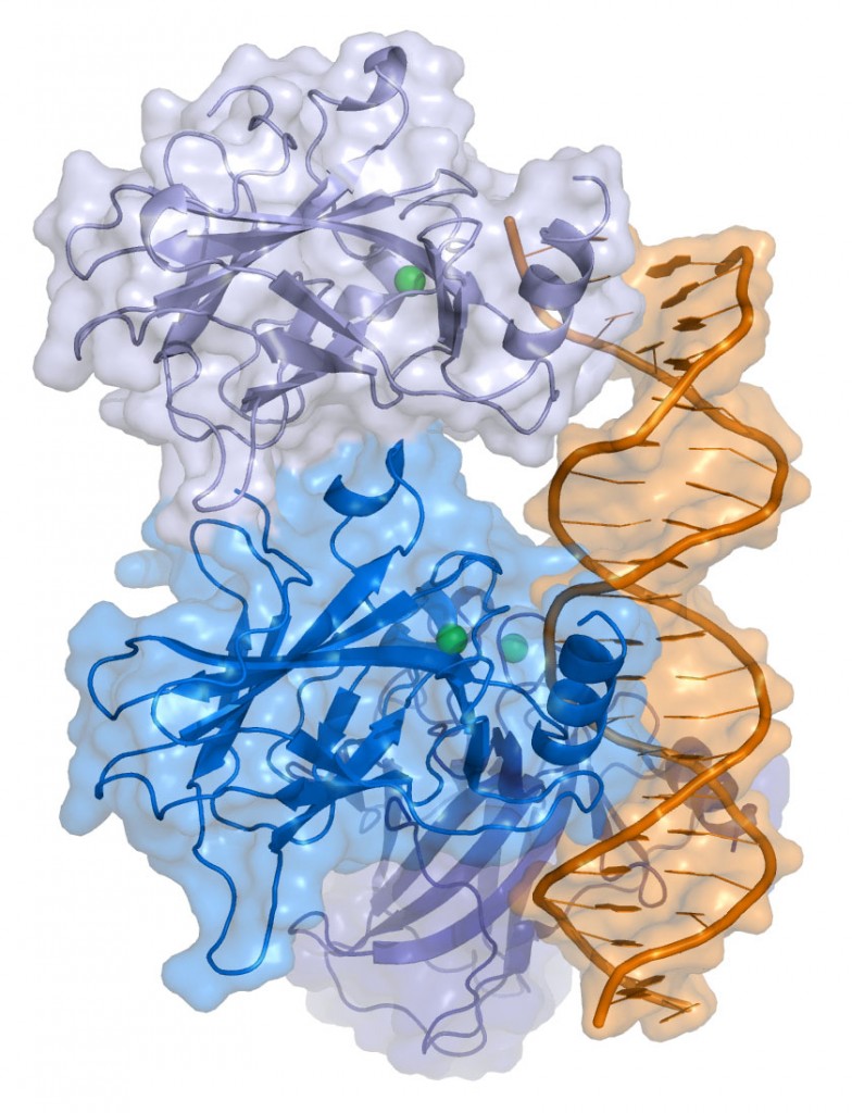
The tumor suppressor p53 is the most frequently mutated gene across all human cancers (Kastenhuber and Lowe, 2017; Laptenko and Prives, 2017). Although implicated in cancer metabolism and genomic integrity via multiple mechanisms, the molecular basis underlying p53-dependent maintenance of genome integrity appears to be fundamentally complex (Aubrey et al., 2018; Brady et al., 2011; Kruiswijk et al., 2015; Valente et al., 2013). Here, we propose crosstalk between p53-driven metabolism and epigenetic control of constitutive heterochromatin as the basis for p53-mediated genomic integrity. Using pancreatic cancer as a model, we show that p53 deficiency leads to a reduction in the universal methyl donor SAM, leaving the cells deprived of defense against epigenetic perturbations. Perturbed p53-deficient cells undergo accumulation of unscheduled R loops, replication stress, and chromosome breakage, leading to genomic instability. R loops originate from massive upregulation of pericentric tandem-repeat transcripts caused by epigenetic instability of constitutive heterochromatin because of inability to preserve H3K9me. Silencing of pericentric tandem-repeat transcripts or excision of unscheduled R loops reinstates genome integrity. Mechanistically, p53 controls expression of genes that influence the methyl donor SAM, including Slc43a2, the transporter for methionine uptake. Thus, cells lacking p53 are metabolically inadequate to preserve global epigenetic integrity upon perturbation, undergoing epigenetic instability of constitutive heterochromatin. Exogenous SAM administration preserves genome integrity even in the absence of p53. Supported by clinical evidence, we propose that metabolic inadequacy is the basis of p53-associated genomic instability.
Results
p53 inactivation correlates with reduced SAM in human pancreatic ductal adenocarcinoma (PDAC)
Oncogenic deregulation has long been associated with metabolic rewiring in cancer (Hirschey et al., 2015). Although the universal methyl donor for DNA and histone methylation, SAM, is acknowledged to represent a critical hub in the metabolism-epigenome interaction, it is unclear whether and how its deregulation participates in cancer pathogenesis. Conducting an immunohistochemistry analysis on a tissue microarray of matched tumor samples of PDAC and normal adjacent tissue, we identified a general reduction in SAM levels in the process of transformation (Figures 1A, 1B , and S1A–S1C). We also revealed that SAM abundance was negatively correlated with p53 mutations in cancer lesions (Figures 1A, 1C, 1D, and S1D). The average SAM level of human pancreatic cancer cell lines carrying p53 inactivating mutations was lower than the average of p53 wild-type (WT) cell lines (Figure 1E). To determine a causative link between p53 and SAM levels, we employed a model system that allowed controlled loss and re-introduction of p53 in an otherwise similar genetic background; this was represented by a panel of mouse PDAC cells derived from tumors that arose in mice carrying pancreas-specific expression of oncogenic Kras (LSL-KRASG12D) and WT Trp53 (KC cells), deletion of Trp53 (KPflC cells, p53−/−), loss of p53 via doxycycline-inducible short hairpin RNA (shRNA) targeting Trp53 (KPshp53 cells) (Morris et al., 2019), or a mutated form of p53 with substitution of arginine with a histidine in codon 270 (KPC270, p53R270H) (Figures S1E and S1F). Levels of SAM were lower in KPflC and KPshp53 cells than in KC cells, but the SAM levels fully recovered upon withdrawal of doxycycline in KPshp53 cells (i.e., reintroduction of p53) (Figure 1F). Thus, inactivation of p53 in PDAC leads to a reduced abundance of SAM.


