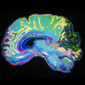
The mammalian brain consists of billions of neurons wired together in various circuits, each one involved in specific physiological functions. To better understand how these different neurons and circuits are associated with mental activities and diseases, researchers are reconstructing detailed, three-dimensional maps of neural networks.
However, 3-D imaging of the mammalian brain is challenging. Light scatters as it travels through layers of tissue, dispersed by a variety of molecules such as water, lipids, and proteins. This reduces image resolution.
One way to improve resolution is to reduce the scattering. Researchers achieve this by first removing water and lipids from tissue. Next, chemicals are introduced that have a refractive index—a measure of how much the molecules bend light that passes through them—in the range of that of proteins. Establishing near-homogenous refractive indices in the molecules that populate the tissue environment allows light rays to converge to improve image resolution. This is the working principle of most tissue clearing methods, which have been used successfully for decades on hard tissues like bone.
Researchers have performed brain tissue clearing with limited success, as the chemicals available were too harsh on delicate neural tissues. In 2013, Karl Deisseroth and his team at Stanford University revolutionized the approach with a hydrogel-based technique called CLARITY. This technique enabled researchers to label neurons in mouse neural tissue with fluorescent markers and then to image an entire mouse brain without sectioning it, while preserving the fluorescence signals.
Since then, tissue clearing methods have continued to improve as researchers have developed chemical mixtures that better preserve tissue architecture and protein structures. The latest techniques are also compatible with fluorescent labels. When they are integrated with advanced tissue processing methods such as automated cell counting, researchers can identify neurons and even intracellular components with ever-improving precision.
Here, The Scientist highlights some of the recent innovations that combine emerging tissue clearing and tissue processing techniques for high-resolution imaging in the brain….
