Abstract
Oocytes form before birth and remain viable for several decades before fertilization1. Although poor oocyte quality accounts for most female fertility problems, little is known about how oocytes maintain cellular fitness, or why their quality eventually declines with age2. Reactive oxygen species (ROS) produced as by-products of mitochondrial activity are associated with lower rates of fertilization and embryo survival3,4,5. Yet, how healthy oocytes balance essential mitochondrial activity with the production of ROS is unknown. Here we show that oocytes evade ROS by remodelling the mitochondrial electron transport chain through elimination of complex I. Combining live-cell imaging and proteomics in human and Xenopus oocytes, we find that early oocytes exhibit greatly reduced levels of complex I. This is accompanied by a highly active mitochondrial unfolded protein response, which is indicative of an imbalanced electron transport chain. Biochemical and functional assays confirm that complex I is neither assembled nor active in early oocytes. Thus, we report a physiological cell type without complex I in animals. Our findings also clarify why patients with complex-I-related hereditary mitochondrial diseases do not experience subfertility. Complex I suppression represents an evolutionarily conserved strategy that allows longevity while maintaining biological activity in long-lived oocytes.
Main
Human primordial oocytes are formed during fetal development and remain dormant in the ovary for up to 50 years. Despite a long period of dormancy, oocytes retain the ability to give rise to a new organism after fertilization. Decline in oocyte fitness is a key contributor to infertility with age2. However, little is known about how oocytes maintain cellular fitness for decades to preserve their developmental potential, complicating efforts to understand the declining oocyte quality in ageing women.
Oocytes remain metabolically active during dormancy6,7, and thus must maintain mitochondrial activity for biosynthesis of essential biomolecules8. Yet, mitochondria are a major source of ROS, generating them as by-products of mitochondrial oxidative metabolism. Although ROS can function as signalling molecules9, at high concentrations ROS promote DNA mutagenesis and are cytotoxic. Indeed, ROS levels are linked to apoptosis and reduced developmental competence in oocytes and embryos3,4,5. However, the mechanisms by which oocytes maintain this delicate balance between mitochondrial activity and ROS production have remained elusive.
Mitochondrial ROS in early oocytes
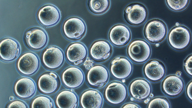
Early human oocytes can be accessed only through invasive surgery into ovaries. Therefore, biochemical investigations into oocyte biology have historically been hindered by severe sample limitations. As a consequence, mitochondrial activity in primordial oocytes remains largely unstudied. Here we overcome challenges imposed by human oocytes by utilizing an improved human oocyte isolation protocol recently developed in our laboratory6, which we combine with a comparative evolutionary approach using more readily available Xenopus stage I oocytes (both referred to as early oocytes hereafter; Extended Data Fig. 1a,b). This approach allowed us to generate hypotheses using multi-species or Xenopus-alone analyses, and subsequently test those hypotheses in human oocytes.
We began our studies by imaging live early human and Xenopus oocytes labelled with various mitochondrial probes that quantify ROS levels. Neither Xenopus nor human early oocytes showed any detectable ROS signal, whereas mitochondria in somatic granulosa cells surrounding the oocytes exhibited ROS and served as positive controls (Fig. 1a–c and Extended Data Fig. 1c–g). ROS induction in oocytes also served as a positive control for live ROS probes (Extended Data Fig. 1h,i)….


