Significance
We demonstrate multiscale phase-contrast X-ray computed tomography (CT) of postmortem human brain tissue. Large tissue volumes can be covered by parallel-beam CT and combined with subcellular detail for selected regions scanned at high magnification. This has been repeated identically for a larger number of individuals, including both Alzheimer’s-diseased patients and a control group. Optimized phase retrieval, followed by automated segmentation based on machine learning, as well as feature identification and classification based on optimal transport theory, indicates a pathway from healthy to pathological structure without prior hypothesis. This study provides a blueprint for studying the cytoarchitecture of the human brain and its alterations associated with neurodegenerative diseases.
Abstract
We have studied the three-dimensional (3D) cytoarchitecture of the human hippocampus in neuropathologically healthy and Alzheimer’s disease (AD) individuals, based on phase-contrast X-ray computed tomography of postmortem human tissue punch biopsies. In view of recent findings suggesting a nuclear origin of AD, we target in particular the nuclear structure of the dentate gyrus (DG) granule cells. Tissue samples of 20 individuals were scanned and evaluated using a highly automated approach of measurement and analysis, combining multiscale recordings, optimized phase retrieval, segmentation by machine learning, representation of structural properties in a feature space, and classification based on the theory of optimal transport. Accordingly, we find that the prototypical transformation between a structure representing healthy granule cells and the pathological state involves a decrease in the volume of granule cell nuclei, as well as an increase in the electron density and its spatial heterogeneity. The latter can be explained by a higher ratio of heterochromatin to euchromatin. Similarly, many other structural properties can be derived from the data, reflecting both the natural polydispersity of the hippocampal cytoarchitecture between different individuals in the physiological context and the structural effects associated with AD pathology.
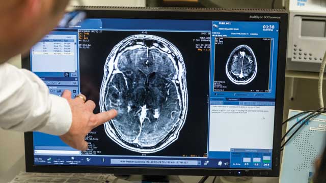
Brain mappings of the cyto- and myeloarchitecture in larger brain areas performed postmortem are required to advance our understanding of the human brain in quantitative terms. Beyond refinements of a brain atlas, they are also essential for later integration of in vivo functional observations with high-resolution structural data (1⇓–3). Mapping the brain, however, requires additional imaging approaches, which can visualize and quantify the three-dimensional (3D) architectonics, including data from more than a single individual (2). The potential of phase-contrast X-ray tomography also known as phase-contrast computed tomography (PC-CT) for 3D brain imaging has been recently demonstrated, both for small animals models (4⇓⇓⇓–8) and the human brain (9⇓⇓–12). Since the entire 3D architecture on all scales is relevant for physiological functions and pathological mechanisms, multiscale implementations of PC-CT (13) are particularly suitable for brain mapping.
Complementary to genomics, proteomics, and metabolics, structural data are also required to unravel mechanisms of neurodegenerative diseases. Such data must be comprehensive (large patient and control groups), quantitative, and fully digital; amenable to advanced analysis including deep learning; and intrinsically three-dimensional. Alzheimer’s disease (AD) is a case in point: Evidence for morphological changes in the hippocampus upon aging and disease can be found already in vivo with MRI. To interpret such data based on a reference model, a 3D probabilistic atlas of the hippocampus was put forward in ref. 3, combining postmortem MRI with histology. The authors concluded that, to test the hypothesis of differential involvement of hippocampal subfields in AD, a “more granular study” of the hippocampus in aging and disease would be required and hence higher-resolution and truly 3D data.
To this end, we here present an advanced and multiscale implementation of PC-CT in combination with automated segmentation and statistical analysis of morphological features. In this way, a much-needed complement to conventional 2D histology is provided, sparing sample sectioning and staining. The signal is generated by the spatial variation of the real-valued part of the X-ray index of refraction n=1−δ+iβn=1−δ+iβ , with δ being proportional to the electron density. Importantly, the advantage of PC-CT derives from the real-valued decrement being significantly larger than the imaginary, absorption-accounting component β; i.e., δ/β≈103δ/β≈103 in the hard X-ray regime. Image contrast is then efficiently formed by free-space propagation, i.e., self-interference of a partially coherent beam behind the object. The fact that this does not require any additional optics between the object and the detector provides a benefit both for dose efficiency and for resolution. Several PC-CT studies have already targeted hippocampal cytoarchitecture in transgenic mouse models for AD (7, 14⇓⇓–17), which exhibit considerable contrast for a typical hallmark associated with this disease, namely β-amyloid plaques. In a recent study, we could also demonstrate the potential of PC-CT on paraffin-embedded hippocampal human tissue affected by AD and evaluate its capability to visualize different pathologies, including plaques, depletion of neurons, or possible recruitment of microglia to affected sites (12).
In this work, we study the 3D cytoarchitecture of the human hippocampus, which serves the formation of declarative long-term memory, i.e., remote episodic or remote semantic memory, but may also affect recent memory, emotions, and vegetative functions (18). Pathologically, the hippocampus is one of the regions first affected in AD (19). As we show here, the throughput of PC-CT measurement, reconstruction, segmentation, and analysis is sufficiently high to treat data from a larger pool, here consisting of postmortem paraffin-embedded tissue blocks of several individuals, both of an AD and of a control group (CTRL), categorized by neuropathological assessment based on the National Institute of Aging – Alzheimer’s Association (NIA-AA)–recommended ABC staging (20, 21). We specifically target the dentate gyrus (DG) and its AD-caused structural alterations. As recently shown, hippocampal neurogenesis and plasticity of the entire hippocampal circuitry are linked to the DG and are found to sharply drop in AD (21). Further, we deliberately do not focus on plaques and tangles in AD, which have already been targeted by a high number of studies, but address in particular the nuclear structure of the DG neurons, since recent evidence points to a nuclear origin of AD (22) including chromatin structures (23). In addition, we also include 3D imaging examples of other parts and structures of the hippocampus and provide the corresponding statistical analysis.
Fig. 1 shows a schematic of the hippocampus that is embedded in both left and right temporal lobes of the cerebral cortex, as a part of the limbic system. In Fig. 1A, the hippocampus is sketched in the sagittal plane, where it forms an elongated structure of about 4 to 5.2 cm in length (24). In Fig. 1B, the frontal plane is shown, in which the appearance is often denoted as snail shaped. Its characteristic functional units are shown in Fig. 1C, most prominently including the cornu ammonis (CA) and the DG, which is a dense zone of granule cells. In the polysynaptic signal pathway relevant in semantic memory formation, input signals from the entorhinal cortex (EC) reach first the DG, which is composed of elliptically shaped granular cells with millimeter-long dendrites. Connected through mossy fibers, information is further processed in the CA, whose neurons are characterized by their pyramidal-shaped bodies. The compartmentalization of the CA with commonly attributed subregions CA1 to -4 is not entirely standardized. The information exits the hippocampus to the inferior temporal cortex, the temporal pole, and the prefrontal cortex, constituting the gray matter (GM). There are further pathways of information processing, involving myelinated tracts in the white matter (WM) that link the hippocampus to further brain regions. The physiological relevance of the hippocampus, with respect to several important signal pathways and its pivotal role in memory function and neurodegenerative diseases, in particular in AD, underpins the necessity to study its 3D structure with cellular and subcellular resolution….


