Abstract
Preclinical studies of primary cancer cells are typically done after tumors are removed from patients or animals at ambient atmospheric oxygen (O2, ~21%). However, O2 concentrations in organs are in the ~3 to 10% range, with most tumors in a hypoxic or 1 to 2% O2 environment in vivo. Although effects of O2 tension on tumor cell characteristics in vitro have been studied, these studies are done only after tumors are first collected and processed in ambient air. Similarly, sensitivity of primary cancer cells to anticancer agents is routinely examined at ambient O2. Here, we demonstrate that tumors collected, processed, and propagated at physiologic O2 compared to ambient air display distinct differences in key signaling networks including LGR5/WNT, YAP, and NRF2/KEAP1, nuclear reactive oxygen species, alternative splicing, and sensitivity to targeted therapies. Therefore, evaluating cancer cells under physioxia could more closely recapitulate their physiopathologic status in the in vivo microenvironment.
INTRODUCTION
The past two decades witnessed substantial advances in development of targeted therapies against pathways altered in cancer cells (1). These agents act against specific cellular dependencies, most often pathways that have undergone oncogenic dysregulation. These advances led to significant improvements in outcome in select groups of patients. Examples include patients with lung cancer with specific mutations in epidermal growth factor receptor (EGFR) who benefit from EGFR-targeted therapies (1). Similar advances have been achieved with immune checkpoint inhibitor therapies (2). While these two are major success stories, many more targeted therapies were less effective in clinic despite promising preclinical data. The best example is PIK3CA (phosphatidylinositol-4,5-bisphosphate 3-kinase catalytic subunit alpha)-targeted therapies. PIK3CA, which is mutated in ~30% of all cancer (the second most common mutation after TP53), is part of the major growth factor–activated signaling networks involved in cell proliferation, survival, and drug resistance. Many drugs targeting PIK3CA failed in clinic, and responses observed with a few tumors did not correlate with PIK3CA mutation status (3). Various mechanisms of resistance to these targeted therapies have been proposed. A convergence-based framework for cancer drug resistance was recently proposed to aid in discovery of new classes of drugs that can overcome resistance (4). Three major mechanisms of resistance described include pathway reactivation, pathway bypass, and pathway indifference. Pathway reactivation involves alterations in drug target as well as upstream, parallel, and downstream effectors of the signaling network targeted by the drug. For example, insulin feedback loop induced by phosphatidylinositol 3-kinase (PI3K) inhibitors reactivates the PI3K-mTOR (mammalian target of rapamycin) signaling axis, thus compromising the effectiveness of PI3K inhibitors. In cases of pathway bypass, the cancer cell finds alternative ways to reengage more downstream oncogenic output. Pathway bypass is often observed in PI3K inhibitor therapies where PIM (proviral integration site for moloney murine leukemia virus) kinases converge onto mTOR pathway (3). Pathway indifference is a situation in which cancer cells acquire an alternative cell state such as mesenchymal state, which permits cancer cells to escape the effects of targeted therapies. Cancer stem cells (CSCs) are typically resistant to many therapies including targeted therapies, and CSC phenotype often overlaps with mesenchymal state (5). Thus, cancers enriched for CSCs may use pathway indifference to become resistant to drugs. In addition, a clear understanding of multifactorial targeted therapy resistance mechanisms is needed to develop effective durable responses to treatment.Most preclinical studies of cancer models including CSC characterization are done at ambient atmospheric oxygen (O2) of ~21%, while physiologic O2 concentrations vary between organs, usually ranging from 3 to 10% (6). O2 concentrations at 5, 2, 1, and 0.4% are defined as physioxia, physiologic hypoxia, pathologic hypoxia, and radiobiological hypoxia, respectively (6). While median O2 concentration in the normal breast is 6.8%, it is 1.3% in breast tumors. This range in O2 concentrations has been described for organs such as brain, liver, head and neck, lung, pancreas, and prostate and for tumors originating in these organs (6). While the effect of O2 concentration on tumor cell characteristics has been studied for a number of years through comparative analyses of cells grown under hypoxia (1%), an O2 concentration required for classic hypoxia-inducible factor (HIF) pathway activation, and ambient air, it was not recognized until recently that exposure of cells to ambient air for even minutes significantly alters the biology and enhances the differentiation of stem cells, effects noted with stem cells in young and old (7, 8). Our group demonstrated that hematopoietic stem cells (HSCs) collected and processed at ambient air undergo extensive differentiation and demonstrate lower engrafting capacity compared to HSCs collected and processed at physioxia (collection at 3% O2 and growth under 5% O2) (8). Exposure of HSCs to ambient air caused extra-physiologic oxygen shock/stress (EPHOSS), which enhanced differentiation of HSCs to hematopoietic progenitor cells.The goal of this study was to determine how collection and processing of tumor tissues under physioxia and ambient air affect CSC characteristics, cellular signaling networks, and sensitivity to therapies. We used two transgenic mammary tumor models and ascites fluids of patients experiencing ovarian and other cancers that metastasize into the peritoneal cavity as model systems. Colon tissues from mice collected and processed at physioxia and ambient air were also analyzed as controls. In every case, tissue was harvested at physioxia, minced, split into two, and then processed at physioxia and ambient air to ensure that comparison is between two fractions of the same tissue but differ only with respect to immediate and subsequent O2 exposure. We demonstrate distinct tumor biology in cells collected and processed under physioxia compared to those collected at 3% O2 and then processed in ambient air and propose that characterizing tumors at physioxia is critical to identify physiologically/pathologically relevant signaling networks and effective responsiveness to drugs that correctly recognize their in vivo status.
RESULTS
Tumor cells display distinct CSC marker profiles under physioxia collection/processing compared to processing under ambient air
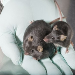
To determine whether continued exposure of tumor cells under physioxia during their collection and processing versus ambient air influences CSC marker profiles, we harvested mammary tumors from MMTV-PyMT (mouse mammary tumor virus-polyomavirus middle tumor-antigen) and MMTV-Her2/Neu+ (mouse mammary tumor virus-human epidermal growth factor receptor 2/Neu+) mice under 3% O2, minced, and divided the tumors into two fractions. Gene expression patterns in mammary tumors of MMTV-PyMT and MMTV-Her2/Neu+ transgenic mice represent Luminal B and HER2–amplified breast cancer subtypes, respectively (9). One fraction was kept at 3% O2, and the other fraction was placed at ambient air for 1 hour. We had previously demonstrated using mouse bone marrow cells that results were similar in terms of collecting cells at ambient air or placing them in ambient air for 1 hour after collecting at 3% O2 (8). After 1 hour of exposure, samples were processed at 3% O2 and ambient air, and cells were either processed immediately for CSC marker profiles, reinjected into animals, or cultured at 5% O2 versus 21% O2. Cells collected, processed, and grown at 3 to 5% O2 are referred to as physioxia and those at 21% as ambient air from here onward. We selected a 1-hour time point because it typically takes at least 1 hour for tumor tissue from surgery to go to pathology and then to research laboratories for functional studies. Samples were processed, stained, fixed, and subjected to flow cytometry for cell surface marker profiling. The cells derived from tumors were characterized in four stages as indicated in Fig. 1A. We selected LGR5 (leucine-rich repeat-containing G-protein coupled receptor 5) and TSPAN8 (tetraspanin 8) as stem cell markers of mammary tumor cells for the following reasons. LGR5-expressing cells are sufficient for postnatal mammary gland organogenesis, and LGR5 is a marker of fetal mammary stem cells (10). In addition, LGR5 is a functional biomarker for CSCs that imparts proliferative and self-renewal capacity to stem cells and has the potential to produce reactive oxygen species (ROS) (11). TSPAN8 is a marker of CSCs and activates sonic Hedgehog signaling (12).FIG. 1…


