Abstract
This study introduces a flexible, adhesive-integrated electrode array that was developed to enable non-invasive monitoring of cervical nerve activity. The device uses silver-silver chloride as the electrode material of choice and combines it with an electrode array consisting of a customized biopotential data acquisition unit and integrated graphical user interface (GUI) for visualization of real-time monitoring. Preliminary testing demonstrated this electrode design can achieve a high signal to noise ratio during cervical neural recordings. To demonstrate the capability of the surface electrodes to detect changes in cervical neuronal activity, the cold-pressor test (CPT) and a timed respiratory challenge were employed as stressors to the autonomic nervous system. This sensor system recording, a new technique, was termed Cervical Electroneurography (CEN). By applying a custom spike sorting algorithm to the electrode measurements, neural activity was classified in two ways: (1) pre-to-post CPT, and (2) during a timed respiratory challenge. Unique to this work: (1) rostral to caudal channel position-specific (cephalad to caudal) firing patterns and (2) cross challenge biotype-specific change in average CEN firing, were observed with both CPT and the timed respiratory challenge. Future work is planned to develop an ambulatory CEN recording device that could provide immediate notification of autonomic nervous system activity changes that might indicate autonomic dysregulation in healthy subjects and clinical disease states.
Introduction
The autonomic nervous system (ANS) links the central nervous system (CNS; brain and spinal cord) with peripheral organ systems, including the integumentary (sweat glands), circulatory (heart, blood vessels), digestive (gastrointestinal tract glands and sphincters, kidney, liver, salivary glands), endocrine (adrenal glands), reproductive (uterus, genitals), respiratory (bronchiole smooth muscles), urinary (sphincters), and visual (pupil dilator and ciliary muscles) systems1,2,3. The ANS is colloquially divided into two main divisions: the sympathetic and parasympathetic nervous systems. However, both branches continuously coordinate through concerted feedback mechanisms to carefully control peripheral organ systems4,5. A large body of empirical evidence suggests that autonomic nervous system imbalance is associated with various pathological conditions that can include heterogeneous disease states, such as diabetic autonomic neuropathy, hyperhidrosis, orthostatic intolerance/postural tachycardia syndrome, pure autonomic failure, autonomic dysreflexia, Takotsubo cardiomyopathy, and vasovagal syncope, and it also contributes to pathophysiology associated with autoimmune inflammatory disorders such as Rheumatoid Arthritis6,7. Moreover, mental health disorders (e.g., Post-traumatic Stress Disorder (PTSD) and Major Depression Disorder) regularly exhibit circadian autonomic dysregulation, with heightened sympathetic and concomitant low parasympathetic drive most commonly reported8,9,10,11,12.
The human cervical spine (neck) is the site of a confluence of autonomic neural structures that are in close proximity to each other, including the major parasympathetic neuronal output transmitted by the vagus nerve13,14. The vagus nerve communicates directly to the visual, heart, respiratory, and digestive systems, and the major sympathetic neuronal output transmitted by the middle and superior cervical ganglion is located approximately 1–2 cm deep to the vagus nerve15,16. The sympathetic ganglion, carotid body, and the vagus nerve are all localized within the carotid artery sheath, and, potentially due to this close proximity, sympathetic fibers have been observed in vagus nerve fascicles (called hitch-hikers), which further indicates multi-sourced neuronal signaling at this cervical level17. The superior cervical ganglion and the thoracic sympathetic ganglion output directly to the integumentary, visual, circulatory, respiratory, and digestive organ systems. Given the immense peripheral organ system control generated from cervical autonomic neuronal structures found within the superficial cervical neck18, decoding these signals to understand the role they play in health and disease could have significant impact on a host of conditions. Prior preclinical work demonstrate resting vagus nerve action potential recording with: (1) cuff electrodes in anesthetized rats during inflammatory cytokine injection19, (2) carbon nanotube yarn electrodes in anesthetized rats during chronic recording20, (3) multi-electrode recording cuff electrodes in regularly breathing anesthetized pigs21, (4) upon stimulation evoked compound action potentials22, and (5) inserting tungsten microelectrodes into the vagus nerve through ultrasound guidance in awake humans23. Further, recent work demonstrate vagus nerve action potentials uniformly synchronize with the respiratory cycle in porcine models24,25 and in one recent human microelectrode study23, although synchronized respiratory and cardiac activity may reflect recording modality deployed26. Other preclinical work measured (via cuff electrode) superior cervical ganglion activity with hypertensive stress tests, i.e., injection of adrenaline27,28 or during painful stimuli29. These studies uniformly demonstrate immediate (within seconds) change in cervical sympathetic neuronal (superior cervical ganglion) activity with each challenge27,28,29. However, the invasive implantation and the risk and complications associated with acute and chronic surgical cuff electrodes have likely precluded, to date, any reported human cervical vagus nerve, carotid body, or superior cervical ganglion recording with validated stress models.
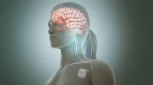
To enable recording with human autonomic stress models, a noninvasive, adhesive-integrated and skin conformal silver-silver chloride electrode array was developed that is capable of conformal positioning over the human left superior anterior cervical area overlying multiple neural structures (i.e., the vagus nerve and its branches, trigeminal nerve branches, sympathetic chain and its branches, the hypoglossal and glossopharyngeal nerves, as well as muscle and dermal sympathetic nerves), and its ability to monitor cervical nerve activity was tested using two widely used and validated stress tests, the cold pressor stress test (CPT) and a timed respiratory challenge. CPT is performed by immersing the hand into a container filled with ice water, which is known to trigger a sympathetic reaction that involves blood vessel constriction and, thus, an increase in blood pressure30,31. It also increases the reflexive modulatory vagal tone by activating multiple brain stem nuclei that coordinate afferent and efferent vagus nerve signaling32,33. The effect of CPT—namely pain—on heart rate is bimodal: subjects routinely demonstrated either an increase or a decrease34,35. Likewise, in timed respiratory challenge studies, muscle sympathetic nerve activity was bimodal; subjects either increased or decreased muscle sympathetic nerve firing36. To date, there is a paucity of inter-challenge analysis within subject physiological measures in response to CPT and timed respiratory challenge. To fill this gap, the newly developed flexible adhesive electrode array was deployed for non-invasive cervical electroneurography (CEN) during a sequential CPT and timed respiratory challenge. Multi-stress challenge CEN measures could help to further disambiguate human autonomic biotypes amongst healthy and disease states in several ways: (1) by facilitating the development of biomarkers of response to pharmacologic and or neuromodulation therapies, (2) enabling the prediction of inflammatory response to pain, and (3) aiding differentiation of sterile vs. non-sterile inflammation.
Results
The custom surface electrode array that was tested is adhesive integrated and flexible, so that it can be non-invasively attached to a subject’s anterior cervical neck. The electrode array was placed lateral to the trachea and medial to the sternocleidomastoid for this study. Silver-silver chloride was utilized as the material of choice for the custom electrodes (Fig. 1) in tandem with a customized biopotential data acquisition unit (Fig. 2c). The custom electrode array allowed subjects to freely move (lateral rotation as well as forward, reverse, and lateral flexion and extension) without distorting the adhesiveness and robustness of the physical structure of the electrode array (Fig. 2a,b)37. The impedance between the custom surface electrode array and the skin was maintained at 1.6 kΩ or less over a 2-h period of testing time; whereas consistent impedance maintenance over a 2-h period is considered ideal for electrical physiological recording38. With consistent low impedance we observed a concordant decrease in power line noise and its harmonics39,40.
Cervical electroneurography recordings were carried out with four electrode “channels” positioned rostral to caudal to evaluate cervical signal: (1) pre-to-post CPT and (2) during a timed respiratory challenge. All channels were run through a spike sorting algorithm to identify putative action potentials or nerve firing patterns associated with different nerve branches. Instantaneous heart rate (beats per minute) at each heartbeat was derived by dividing the 60 (s/min) by the extracted RR intervals calculated from our ECG recordings.
In a sample subject, the change in cervical neural firing was observed to coordinately increase with onset of CPT; the cervical neural firing was extrapolated from the spike sorting algorithm (Fig. 3a). Simultaneous recordings from Channel #1 (rostral overlying nodose ganglion, C2/3 cervical dermatome, and auriculotemporal branch of the trigeminal nerve) and heart rate were compiled (Fig. 3a). Amongst an array of clusters, responsive clusters were identified. Responsive clusters were defined as clustered groups that significantly increased in firing (by greater than 2 standard deviations) during CPT (for at least 40% of the stress challenge) compared to pre-CPT baseline activity….


